Farm March Newsletter 2026

Dedicated farm line – 01626 357776

Follow us on Facebook

https://www.facebook.com/Axiomfarmvetlab/

This newsletter is sent by e-mail to each vet practice but if you would like a copy sent to your individual e-mail account please contact us at dsfarm@axiomvetlab.co.uk and we can add you to our circulation list.

Would you like to send the Axiom farm vets photos of PMEs to allow us to assist with sample selection and diagnosis? Please send photos by WhatsApp to 07944 649222.

You may find this is a more efficient way of making requests than phoning the farm team, saving you time in your busy day. Our farm team also find it a more efficient way of dealing with your requests.
The email address for test requests is: dsfarm@axiomvetlab.co.uk 

Axiom Farm Vets

If you wish to speak to a particular farm vet about one of your cases, you can consult the table below to see when our vets are usually available. A duty farm veterinary advisor is always around on a Saturday morning from 9am-12pm.

Farm Vets

BTV testing guidance

We wish to remind vets that as a commercial laboratory we are unable to offer any Bluetongue testing either on blood samples or abortion material. If we receive samples from cases in which Bluetongue is suspected, we are required to safely dispose of the samples and cannot provide any further testing. We are always happy to discuss cases over the phone before submission of samples. Clinical suspicion of BTV infection must be reported to APHA. Further information on investigating poor reproductive performance in cattle and sheep during Bluetongue outbreaks can be found at the following link:

BTV Web Link

High Pooled Inorganic Iodine results

High Pooled Inorganic Iodine results

Unexpected very high results in the Pooled Inorganic Iodine (PII) test do occur from time to time. These elevated results are not straightforward to interpret. It is worth double checking for hidden iodine sources. Mineral licks or buckets may have a high iodine content, and some individuals may take considerably more than the expected daily intake raising the PII level of the pooled sample. Kelp or seaweed-based supplements are usually high in iodine. Disinfectant used in parlour hygiene or for calf/ lamb feeding equipment or for treatment of navels may contain iodophors. Some water sources, particularly borehole water is known to contain high level of trace minerals. Unexpected high values tend to occur without clinical signs. Therefore, iodine toxicity is rarely identified but when it does occur signs can be vague and include persistent coughing, naso-ocular discharge, inappetence, reduced growth rates and dermatitis/ alopecia. Long term high iodine intakes can reduce iodine uptake by the thyroid gland and can cause goitre. Various studies have shown that lambs born to ewes with high iodine intakes had poorer colostral absorption.
If no source of excessive iodine can be identified, then the result may need to be confirmed by resampling. Please let one of our vets know if you plan to recheck a PII result so that it is flagged as a herd/flock with a previous high result and can be interpreted with this in mind.

Foetal stomach contents

To maximise the chance of a diagnosis in abortion cases, foetal stomach content (FSC) is one of the most useful samples we can receive. It is used primarily to identify bacteriological and fungal causes of abortion. It is an important component of our abortion PCR package (Test code: FABORT) where it is used in a ‘pool’ along with fresh foetal brain and liver tissue. In a freshly aborted foetus, the abomasal contents will be relatively protected from environmental contamination as opposed to, for example, placental tissue which tends to be heavily contaminated. When sampling FSC the risk of contamination can be minimised by using a sterile vacutainer and needle punctured though the stomach wall to sample the fluid inside. Where insufficient fluid is present then an alternative is to nick the stomach wall with a clean scalpel and introduce a swab (charcoal or other swab with transport media suitable for bacteriology) through the wall to sample the contents. FSC is only suitable for testing in abortion cases and cases at term where the animal was known to have been stillborn. Stomach contents from animals that have been alive for several hours/ days are not suitable for testing as they may have received milk/milk replacers or attempted to suckle contaminating of the stomach contents and leading to less than useful results e.g detection of the agent in milk/colostrum.

Haemonchus contortus test

We have validated a new method for differentiation of Haemonchus species from other Strongyle eggs in faecal samples. Haemonchus contortus speciation is now performed using the OvaCyte system and this method compares well with the PNA (peanut agglutinin test) * and offers a reduced turnaround time. Specificity has been estimated to be around 90%**. If a positive result is very unexpected (e.g. from a flock with no known history of Haemonchosis or a closantel post-drench check etc) it is important to be aware that the risk of a false positive cannot be entirely excluded. False positive risk may be higher in samples with a low total strongyle count. The PNA test continues to be available to further investigate an unexpected positive result if required.

*Correction from February issue: ‘Peanut agglutination test’ corrected to ‘Peanut agglutinin test’ **Elghryani N, Lahan G, Bor Gohain J, McOwan T and de Waal T (2025) Comparison of OvaCyte™ Speciation and PNA staining for the detection of Haemonchus contortus in ovine faecal samples. Front. Vet. Sci. 12:1688644. doi: 10.3389/fvets.2025.1688644

Manganese testing

We have taken the decision to suspend our referral test for manganese on bloods for the foreseeable future, due to concern over reliability of recent results and inconsistencies in suggested reference intervals in the literature. Whole blood, plasma and serum manganese levels are known to be highly variable and generally considered to be a poor indicator of manganese status in the animal. Liver manganese levels are also variable but may be a more reliable indicator of status. Where manganese deficiency is suspected due to the birth of calves with appearance consistent with congenital joint laxity and dwarfism (CJLD) consider increasing manganese supplementation in the following season. This should be done as part of a range of measures which can include increasing level of non-ensiled feed to at least 25% Dry Matter intake*, providing small amounts of supplementary concentrates and ensuring minimum National Research Council (NRC) mineral guidelines are met. If you are considering sending blood samples for manganese testing, please feel free to contact us to discuss your case.
Correction from February issue: Percentage DMI altered from 30% to 25% to reflect current recommendations.

Interesting cases in January

Cattle
Abortions/mastitis

  • Salmonella Dublin, Campylobacter fetus, Listeria monocytogenes, Bacillus licheniformis and Trueperella pyogenes were isolated from foetal stomach contents in cases of bovine abortion this month.
  • A heavy and pure growth of the yeast Candida galbrata was isolated in fungal cultures of foetal stomach contents in a case of bovine abortion. Candida galbrata is a recognised pathogen in humans and cattle and as it was isolated as heavy and pure growth in cultures it was of likely significance. Abortions due to Candida species are uncommon in cattle. It was suggested that if further cases occur on this farm to consider submitting placental samples for histopathology as placental lesions may be present in mycotic abortions.
  • Foetal fluids were positive for BVD antigen in at least five cases of bovine abortion this month. In two of these cases a significant bacterial cause of abortion was also identified in cultures.
  • Evidence of Neospora caninum infection was detected by PCR of foetal tissues in several cases and in at least one case by detection of antibodies in foetal fluid. One of the PCR positive cases underwent histopathology on brain tissue which confirmed Neospora infection in the central nervous system and confirmed Neospora as the likely cause of abortion.
  • Streptococcus canis was isolated from milk submitted for mastitis cultures. Streptococcus canis is a rare cause of bovine mastitis. Typically, it is associated with sporadic cases but occasionally it may cause outbreaks of mastitis due to cow-to-cow spread.

Respiratory disease

  • IBR was detected in nasal swabs from a two- year-old fattening heifer with signs of respiratory distress, increased upper airway noise and halitosis. Other cattle in the group had been showing similar signs and two had died. Necrotising tracheitis was observed at postmortem of one animal. In another IBR case a recently calved cow showed signs of pyrexia, increased upper respiratory tract noise and increased respiratory rate. IBR was detected in nasal swabs by PCR and a bulk tank IBRgE serology was strongly positive for antibody to wild type IBR. On a third farm convalescent blood samples were received from animals which had shown signs of clinical IBR several weeks before. Of three animals tested two had seroconverted from negative to strong positive in the IBR antibody test and the third had gone from a low positive result to a high antibody level, confirming recent exposure to IBR in all three animals. At least 19 further cases of IBR were detected by PCR in January.
  • A group of calves underwent thoracic ultrasound on arrival at a rearing unit, and scans were deemed to be clear of lung lesions and the calves appeared in good health. Three weeks later re-scanning identified significant lung pathology as present indicating that the disease had occurred post arrival. Pooled PCR testing of four nasal swabs from affected calves detected Coronavirus as well as Histophilus somni, Pasteurella multocida and Mannheimia haemolytica. The significance of these findings on nasal swabs can be difficult to interpret. Bovine coronavirus has been suggested in some studies to be involved in BRD, but it can also be found in healthy cattle. The findings of Histophilus somni, Pasteurella multocida and Mannheimia haemolytica from nasopharyngeal swabs are of uncertain significance as they can be carried in the upper respiratory tract of healthy cattle. However, a study by Doyle (2017) found there was very good correlation between nasal or NP swabs or BAL samples and trans-tracheal washes with regards to Mannheimia haemolytica, Pasteurella multocida and Mycoplasma bovis. Reference : D Doyle, B Credille, T W Lehenbauer, R Berghaus, S S Aly, J Champagne, P Blanchard, B Crossley, L Berghaus, S Cochran, A Woolums. Agreement Among 4 Sampling Methods to Identify Respiratory Pathogens in Dairy Calves with Acute Bovine Respiratory Disease. J Vet Intern Med. 2017 May; 31(3):954-959.
  • There were significant rising antibody levels to RSV on paired serology of all five tested beef bulls from a group in which there had been a significant respiratory disease outbreak the previous month. RSV was detected by PCR of nasal swabs or lung tissue in at least 43 other cases of respiratory disease in cattle in January.
  • On one farm multiple calves aged around two months were affected with respiratory disease, drooping ears and some had swollen joints. Mycoplasma bovis, Pasteurella multocida and Mannheimia haemolytica were detected by PCR of lung tissue. Histopathology identified chronic fibrous bronchointerstitial pneumonia. As with many cases of bovine respiratory disease findings were complex indicating multiple underlying components. There were changes consistent with Mycoplasma bovis infection but also a fibrinous pneumonia and pleurisy component more typical of pasteurellosis.
  • Klebsiella pneumoniae was isolated in profuse growths from the lung tissue of a Holstein cow which showed signs of respiratory distress three days after calving. At postmortem the lungs appeared discoloured and congested and there was a large amount of serosanguineous fluid present in the thorax. Histopathology of lung revealed moderate-marked, acute, multifocal, necrotising pneumonia with extensive fibrinosuppurative pleuritis and abundant intralesional bacteria. The changes in the lung were most suggestive of septicaemia in this case. K.pneumoniae is a known cause of pneumonia, septicemia and mastitis in cattle.
  • Post-mortem tissues were received from a yearling Holstein-Friesian dairy heifer. The animal showed clinical signs of respiratory disease but failed to improve even with several treatments and died one week later. At this time several others in the group also showed signs of respiratory disease. At postmortem examination diffuse lung pathology was observed. No lungworms were seen in the airways. Respiratory PCR testing of lung tissue detected none of the eight common respiratory pathogens. Histopathology identified extensive, severe, subacute, fibrinonecrotising interstitial pneumonia with fibrinous pleuritis as well as extensive, severe, subacute fibrinous pericarditis. A septicaemia was suggested as a possible cause of the interstitial pneumonia and ruling out salmonellosis with culture or PCR testing was suggested. In addition to the disease processes identified in the lungs, A dense population of round cells was found attached to one section of the pericardium. assisted in identifying. Scattered mast cells and few structures resembling Hassal’s corpuscle were seen throughout suggesting this may have been part of a thymoma. Thymomas are not uncommon in cattle, but most will go unrecognized due to the lack of clinical signs associated with them.

Gastrointestinal disease

  • In one herd thirteen calves had died within 24-48 hours of birth, showing signs of salivation and liquid diarrhoea. A faecal sample from a one-day old calf showing these signs tested positive for F5 (K99) E.coli antigen confirming enterotoxigenic Escherichia coli infection.
  • A fifteen-month-old Angus cross bullock presented for loss of condition and persistent scour. No nematode or fluke eggs were identified on microscopy and testing for BVD antigen and OvHV2 (MCF) by PCR were both negative. Johne’s PCR testing on faeces was positive, supporting a diagnosis of Johne’s disease in this case.
  • Samples were received from a fourteen-month-old bullock which was observed to have scour prior to death. At gross postmortem the gut had a thickened appearance. The strongyle egg count in a faecal sample was 1250epg , confirming a heavy gastrointestinal parasite burden. Salmonella cultures were negative. Histopathology revealed marked chronic ulcerative/necrotising gastroenteritis and colitis with vasculitis, but the underlying cause of the changes was not evident. No viral inclusions were seen to implicate adenovirus and BVD was excluded by PCR testing. It was not clear whether the vasculitis observed was a primary lesion or secondary lesion and it was suggested to also rule out malignant catarrhal fever (MCF) as a cause of vasculopathy. Given the high worm egg count, parasitic gastroenteritis with secondary infection remained a possible cause for the lesions.
  • There were at least eight cases of gastrointestinal disease associated with Salmonella Dublin in January, seven with Salmonella Typhimurium, four with Salmonella Mbandaka and one of Salmonella Give. Salmonella Give is a serotype most associated with poultry but can cause scour and abortions in cattle and in this case was the cause of acute and severe dysentery in a heavily pregnant dairy cow.
  • Fixed samples of jejunum and caecum were received from an eighteen-month-old Galloway heifer which was one of three which had presented as thin and scouring. Necrotic enteritis had been observed at the gross examination. Unfortunately, due to poor preservation the jejunal sample was not useful for diagnostic interpretation but the sample from the caecum showed evidence of a moderate multifocal necrotising typhilitis with evidence of damage to the crypts/ glands but with no obvious underlying cause. Crypt/gland damage may be mediated by toxins, mucosal ischaemia or viral infections such as BVD, coronavirus (winter dysentery) or coccidiosis. Coccidiosis was thought unlikely in an animal of this age, unless immunologically naive due to lack of exposure and heavily exposed to pathogenic coccidia (or immunosuppressed due to, for example, persistent BVD viraemia). Liver analysis identified evidence of copper, selenium, and cobalt deficiency with levels of all trace elements falling below the expected reference intervals. It was not clear what role if any the trace element deficiency may have had in the gastrointestinal disease.

Skin and eye disease

  • Treponema phagedenis, one of the treponeme species associated with digital dermatitis in cattle was detected by PCR testing of a swab from lesions on the medial coronary band of an affected cow.
  • Some animals in a group of eight to twelve-month-old dairy heifers showed signs of skin lesions and intense pruritis. No ectoparasites were seen on microscopy but fungal elements were present. Trichosporon sp. were isolated in dermatophyte cultures confirming a diagnosis of ringworm. Five animals in the group were blood sampled, and all had raised pepsinogen levels consistent with significant abomasal damage due to parasitism. Outbreaks of ringworm in cattle may be associated with concurrent disease (such as parasitic gastroenteritis) and poor immunity.

Systemic and miscellaneous disease

  • Postmortem tissues were received from a three-month-calved primiparous Holstein-Friesian dairy cow which showed sudden signs of discomfort, milk drop and recumbency. Multifocal abscessation of lung, heart, kidney and udder tissue as well as an external abscess (location unknown) were observed at postmortem examination. Heavy growths of Trueperella pyogenes were cultured from both lung and heart abscesses. On histopathology severe and ongoing pathology was observed, with chronic abscessation in the heart and lung. There was marked bacterial endocarditis, and extensive infarcts in the kidney with an underlying chronic nephritis. It was not possible to determine the initiating cause, but embolic spread of bacteria from a focus elsewhere, such as mastitis, metritis, foot abscess or injection site lesion was suggested as a possibility.
  • Postmortem tissues were received from a White Park cow. At gross examination liver abscessation had been observed, multifocal firm and red areas were seen within the lung tissue and there was extensive ulceration in the rumen. On histopathology the lesions in the lung were identified as intravascular fibrin thrombi with intralesional fungal hyphae (fungal emboli) (Figure 1). Given the extensive lesions seen in the rumen, it was considered possible that the spread had occurred from that site. In the liver there was a locally extensive, severe, chronic-active, necrotising hepatitis with intralesional bacteria. The observed bacteria were demonstrated to be gram-negative and Fusobacterium necrophorum was considered the likely cause of this bacterial abscess. Again, the rumen was the most likely site of origin of the bacteria.

Fungal hyphae

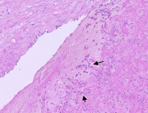
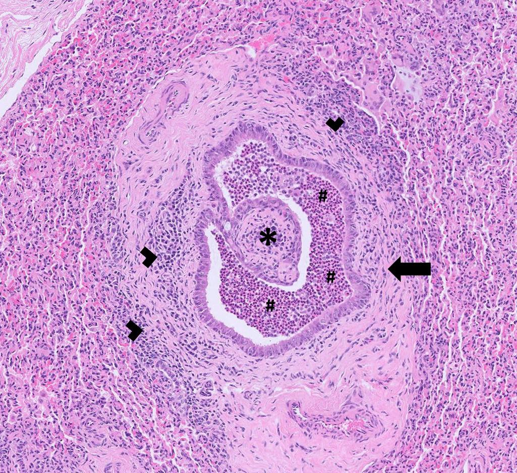
Figure 1 : Fungal hyphae (arrows) seen in a thrombus within a vessel in the lung of a cow

  • Significant cardiac fibrosis was seen on histopathology of heart tissue from a dairy cow which had died suddenly. It was not possible to determine the underlying cause of fibrosis, though a historic bacteremia or toxic insult were thought possible. It was also difficult to know whether the fibrosis could have been associated with the death, but it was considered a possibility if it had triggered a sudden dysrhythmic event.
  • Endocarditis was thought to be the possible cause of leukocytosis (WBCC = 21.4 x10^9, RI = 4-10 x 10^9) in a first lactation dairy heifer which showed signs of inappetence, milk drop and had a heart murmur on auscultation.
  • Formalin fixed brain and spinal cord were received from a six-month-old Aberdeen Angus cross calf which had shown signs of opisthotonus, blindness, nystagmus and paddling movements prior to death. Histopathology of the brain identified severe, subacute, symmetrical, necrotising polioencephalopathy, consistent with a diagnosis of cerebrocortical necrosis (CCN). The lesion distribution and nature were thought to be most suggestive of a thiamine dependent encephalopathy or sulphate toxicity. The former is commonly associated with recent changes in diet resulting in rumen dysbiosis and the production of thiaminase with subsequent vitamin B1 deficiency. Sulphate toxicity is associated with excess intake through concentrate food and/or water.
  • Mucosal disease subsequent to persistent infection with BVD virus, was suspected to be the cause of diarrhoea and mouth ulcers in a calf (age and breed unknown). The animal was positive for BVD in the antigen ELISA test confirming BVD viraemia.
  • Malignant Catarrhal Fever (MCF) due to infection with Ovine herpes virus -2 was diagnosed by PCR testing in at least two cases this month.
  • Lipomatosis (abdominal fat necrosis) was identified as the cause of a mass found adjacent to the large intestine in an eight-year-old dexter cow which had initially presented with signs resembling milk fever. Although initial treatment appeared successful the cow later deteriorated and died. Histopathology identified the mass as necrotic fat, typical of the condition lipomatosis which can occur in any part of the omental, mesenteric or retroperitoneal fat. The aetiology is unclear and although it is most usually an incidental finding, it was noted that it could be the potential cause of gastrointestinal obstruction.
  • Fourteen store calves showed signs of polyarthritis shortly following an outbreak of respiratory disease in the same group. The farm had recently started buying in dairy-cross calves for stores/finishing but also had a Galloway suckler herd on farm. Joint fluid was aspirated from the carpal joint of one of the affected animals and this sample tested positive for Mycoplasma bovis by PCR. Nasopharyngeal swabs taken from the animals with signs of respiratory disease had also tested positive for M.bovis. It was suspected that the pathogen had been introduced into the herd with the bought-in animals as M.bovis infection had not been previously identified in the suckler herd.

Sheep and goats

Abortions, reproductive disease, mastitis

  • At least six cases of abortion associated with Campylobacter fetus were reported. In one flock of 260 Texel ewes, three weeks off lambing, 15 aborted over 10 days. The farm also had a flock of Suffolks, grazing in a different pasture, none of which were affected.
  • Several cases of enzootic abortion were reported, the majority diagnosed on MZN-stained placental smears but also, in one case, Chlamydia abortus PCR. In one positive submission, the flock of 90 ewes were vaccinated against enzootic abortion, and six ewes had aborted but remained clinically well.
  • Salmonella Montevideo was reported as the cause of abortion in one sheep flock. Typically, this serotype is associated with sporadic cases of abortion, but outbreaks of abortion associated with S. Montevideo in southeast Scotland have been linked to scavenging birds, particularly herring gulls.
  • Yersinia pseudotuberculosis was reported as the cause of abortion in a two-year-old ewe. Wild birds and rodents are known to be carriers of Y. pseudotuberculosis. It was advised that, if ewes are being fed in troughs, then, if possible, the troughs should be turned over after they have finished to avoid bird and rodent faecal contamination.

Respiratory disease

  • Post-mortem tissues were received from a two to three-week-old Suffolk ram lamb, one of a number to develop respiratory signs within a few days of life. Fusobacterium necrophorum and E. coli were isolated and histopathology found a large area of necrosis in the lung which was likely the result of F. necrophorum infection, with gram-negative filamentous bacteria seen at the edge of the lesions consistent with the bacterium (Figure 2). Elsewhere in the lung there was suppurative bronchopneumonia and in the airways, there were short gram-negative rods, which could have been consistent with the E. coli isolated or potentially a member of the Pasteurellaceae, although the ewes were vaccinated. F. necrophorum is an opportunistic pathogen commonly found in the environment of farm animals and the route of infection in lambs can include via the navel, ringing or tail docking sites, or through the gastrointestinal tract. There was also a suppurative bronchopneumonia and an
    underlying/concurrent colisepticaemia, or previous/concurrent Pasteurella pneumonia could have been implicated. A review of lambing pen hygiene, colostrum and navel management was suggested.

HE and gram stain

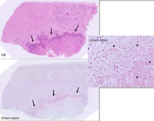
Figure 2 : HE and gram stain sections of lung showing large areas of necrosis (arrows) with gram negative filamentous bacteria (*) confirmed on bacteriology as Fusobacterium necrophorum

  • Postmortem samples were received from an unvaccinated four-week-old Charollais lamb that had died acutely. PME revealed ‘abnormal’ lungs, Mannheimia haemolytica was isolated from lung tissue and histopathology of lung detected an interstitial pneumonia consistent with M. haemolytica septicaemia.
  • In addition to several cases confirmed on serology, Maedi visna was confirmed in a ewe on histopathology. The ewe was barren and developed hind limb ataxia, unilateral weakness and head tilt a couple of hours before dying. Histopathology revealed diffuse, severe, chronic, lymphohistiocytic interstitial pneumonia with smooth muscle hyperplasia and bronchus associated lymphoid hyperplasia, highly suggestive of Maedi (Figure 3). Visna could also have accounted for the neurological signs seen but brain was not submitted for histopathology.

Lung histopathology

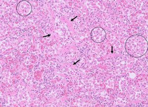
Figure 3 : Lung histopathology from a sheep showing lymphohistiocytic interstitial pneumonia with macrophages in alveoli (circles) and smooth muscle hyperplasia (arrows) suggestive of maedi visna (MV).

  • OPA was confirmed on histopathology in two ill thrifty sheep that were reported as sudden deaths. Animals commonly present with exercise intolerance and ill thrift. However, if a secondary bacterial pneumonia develops, this can often lead to sudden death. In this case there was evidence of mild chronic-active bacterial infection in one animal and a more severe and extensive acute bacterial pneumonia in the other. Likely bacterial pathogens to be involved would include Mannheimia haemolytica, Bibersteinia trehalosi and Pasteurella multocida.

Gastrointestinal disease

  • Parasitic gastroenteritis was reported in sheep and goats, with signs including scour and condition loss. High pepsinogen results consistent with significant abomasal parasitism were also reported in some sheep. In one group of scouring, dying lambs, there was also evidence of nematodiasis.
  • Fasciolosis was confirmed in several sheep flocks on coproantigen ELISA and faecal microscopy, including in two adult babydoll sheep with recurring loose faeces over the course of a month.
  • A total of 101 submissions for Johne’s disease serology and faecal PCR testing from sheep and goats were received, with 9 submissions, including a total of 13 individual samples, testing positive. Cases included one of a group of very thin ewes in a flock which had high worm egg counts and a low scanning percentage, and a three-year-old Alpine goat with scouring and rapid weight loss. One three-year-old Welsh Mountain ewe, one of five emaciated ewes to die in a flock over the last 12 months, was positive for Johne’s on faecal PCR and for liver fluke on coproantigen ELISA, as well as having a strongyle egg count of 900 epg.
  • A Golden Guernsey goat developed acute onset diarrhoea. Worm eggs and coccidial oocysts were seen on in house testing but, despite treatment, the animal died 14 hours later. The only finding on PME was watery gut contents but Type D clostridial enterotoxaemia was suspected on clostridial toxin testing. This was also found in a 10-month-old ewe lamb that died acutely on a second farm, the second to do so in a group of 30, with clostridial toxin testing identifying it as a probable case of pulpy kidney.

Skin and eye disease

  • Sheep scab was confirmed on skin microscopy on at least one occasion and seropositivity for sheep scab was also detected in several flocks with clinical signs including pruritus, wool loss, thickening and scabbing of skin.
  • A recurrence of staphylococcal dermatitis associated with S. aureus was confirmed on culture of a skin swab from a six-year-old pygmy goat with poor skin.
  • Swabs were taken from the eyes of three sheep in a flock that had problems with sore eyes with neovascularisation over a 6-month period. The farmer has treated with cloxacillin eye ointment and long acting oxytetracycline, with resolution in some animals but recurrence in others. Mycoplasma conjunctivae was detected on PCR; this is the most common cause of infectious keratoconjunctivitis in sheep and cases can be difficult to successfully resolve due to failure to eliminate the bacterium from deep within the conjunctival sac. At least two further cases were reported this month.

Systemic and miscellaneous disease

  • A screen of 16 rams in one flock detected four animals seropositive for CLA. Due to the very high specificity of the CLA ELISA used at Axiom, false positives are very rare.
  • A five-year-old-tup was screened on serology for CLA 18 months ago with a negative result but had continued to develop abscesses at various sites. It was seronegative again when screened but an Actinobacillus spp. was isolated in culture consistent with a diagnosis of cutaneous actinobacillosis. Cutaneous trauma is understood to be the route of entry of infection.
  • Among other isolates from suspected CLA lesions this month was Yersinia pseudotuberculosis. This bacterium is a recognised pathogen in small ruminants and has been previously isolated from abscesses and enlarged lymph nodes in sheep.
  • Post-mortem tissues were received from a six-year-old sheep that died after a 24-hour period of lethargy. On PME, there were multifocal swellings of the liver and spleen. Histopathology was consistent with adenocarcinoma of the liver and spleen with hepatic necrosis also present. It was not possible to determine the origin, but this could have been a primary liver tumour, or it may have occurred after metastatic spread of an intestinal adenocarcinoma, which is fairly common in sheep. These cases are usually sporadic.

Camelids

  • High strongyle egg counts consistent with parasitic gastroenteritis were reported in alpacas and Bactrian camels. In two Bactrian camels in a zoological collection, 23% and 28% of strongyle egg counts of 2,032 and 16,400 epg respectively were determined to be Haemonchus sp. by the Ovacyte method. A third camel in the same collection was euthanased following ongoing weight loss, lethargy and recumbency and had a strongyle egg count of greater than 50,000 epg, again likely to be consistent with haemonchosis. On histopathology of liver of this animal, there were multifocal myelolipomas, grossly appearing as white spots. These are benign tumours consisting of collections of adipocytes associated with trabecular bone and these have been identified in numerous exotic species. One previous report in a camel was identified in the literature.
  • Fasciolosis was diagnosed in a recumbent 14-year-old female alpaca on faecal microscopy.
  • Biopsies were received from a growth on the pinna of a 12-year-old male llama. Histopathology found that the mass was comprised of bland spindle cells and a fibroma was the most likely diagnosis.

Other Species

  • Faeces samples were received from a group of seven-week-old pigs from a petting farm with yellow, pasty scour. There had also been one death. F4 (K88) and F41 E. coli and Rotavirus Type A were detected, all of which may have been contributing to the reported scour.
  • Parasitic gastroenteritis was confirmed in a six-year-old pig that was losing weight and had a strongyle egg count of 1300 epg. It was last treated with an anthelmintic in June 2025.
  • Fasciolosis was diagnosed in a reindeer on coproantigen ELISA.
  • Post-mortem tissues were received from a six-month-old, post weaned, male red deer, one of 250 weaned deer that had been housed for approximately three weeks. Four of the deer had become recumbent in the last two weeks, some with respiratory signs, and had died or been euthanased. On PME, fibrin and adhesions were widely present in the thorax, and the lungs were almost fully adhered to body wall. Histopathology showed marked, subacute, fibrinosuppurative and necrotising pleuropneumonia with intralesional gram-negative and gram-positive bacteria. The gram-negative bacteria were possibly members of the Pasteurellaceae and the gram-positive rods were consistent with the Trueperella pyogenes isolated on culture, a common secondary invader.
  • The fourth chicken to die in a flock of around 20 birds was PM’d, revealing peritonitis with fibrin deposits including plaque like adhesions on the serosal surfaces of the gizzard and intestines. A pure heavy growth of E. coli was isolated on aerobic culture of a swab. This may have been a case of egg peritonitis, or the result of an ascending reproductive infection, secondary to air sacculitis, or post-vent trauma.

Deficiencies and toxicities

  • There were at least nine submissions in January in which potentially significant low plasma copper levels were identified in cattle herds. One herd reported previous issues with uterine prolapses in the autumn calving group and wanted to check the nutrition of the next group due to start calving in February. All five tested cows were hypocupraemic and all had low urea levels indicative of low rumen degradable protein intake. Two of the group had raised NEFA levels indicative of excessive fat mobilisation consistent with an energy deficit in the diet. One animal was hypoalbuminaemic. One possible cause of low albumin is long term low protein intake which could have been the case here, but it was suggested that other causes such as Johne’s disease, liver fluke infection or chronic inflammation could also be investigated.
  • Postmortem tissues were received from a six-month-old calf which had shown signs of ill-thrift and later became recumbent and died a short time after. At PME, the kidneys appeared haemorrhagic and bloody peritoneal fluid and urine were observed. The liver had an abnormal orange colour. The copper level in kidney tissue was high at 723umol/kg DM (RI: 141-314umol/kg DM) raising concerns of possible copper toxicity. Histopathology identified hepatic necrosis with intracytoplasmic copper accumulation and examination of kidney showed severe acute tubular degeneration and necrosis. These findings were supportive of a diagnosis of copper toxicity, with evidence pointing toward chronic copper accumulation and hepatopathy followed by acute haemolytic crisis. No evidence of fascioliasis (which could predispose to the acute crisis) was seen in the liver sections submitted. However, a fluke antigen ELISA test on faeces gave a suspicious result of 7.7% (Axiom’s suspicious range = 2-8%). Results classed as suspicious would still have a specificity of >99% if classed as positive in this test. Therefore, it was suggested that liver fluke infection could be a significant factor in this case and further investigation of this in the rest of the group as well as an urgent review of copper levels in the diet was recommended.
  • Formalin fixed liver samples were received from a weaned dairy calf. The calves were receiving 2kg/head of concentrates along with round bale silage which appeared to contain large amounts of ragwort (Senecio jacobaea). Three animals from the group had died and a further two calves were sick and recumbent. Due to poor prognosis these were euthanased and underwent postmortem examination. Liver sections were examined and evidence was found of centrilobular to midzonal, acute, moderate, hepatic necrosis with moderate sinusoidal leucocytosis. There was no evidence of copper accumulation with Rhodanine special stain. Possible causes of the hepatic necrosis include terminal hypoxia (this is usually worse in animals that have died slowly, are anaemic, or have cardiac disease) or a toxic insult. These changes are therefore non-specific, but acute ragwort toxicity was considered likely given the clinical history.
  • A blood sample was submitted from a seven-year-old Holstein- Friesian dairy cow which had suffered a prolapse after calving. The farm was reported to have had recent increases in milk fever cases, uterine prolapses and endometritis. This cow had a critically low calcium level of <1mmol/l (RI 2.00-3.00) as well as severe hypophosphataemia with an inorganic phosphate level of 0.33mmol/l 9 (RI 1.1-2.5mmol/l).
  • Blood samples were received from three of a group of dairy-cross growing bulls, with locomotor disturbances (stiffness, going off back legs) and a high incidence of long bone fractures. One animal tested had evidence of hypocalcaemia and hypophosphataemia. Blood levels of the macrominerals do not always reflect levels in bone due to tight homeostatic regulation so low calcium/phosphorus levels in bone could not be ruled out in the other two animals. One of the three cattle had a low GSH-Px result consistent with selenium deficiency and the other two were slightly above the cut-off value of >30U/ml RBC. All three tested cattle also had a vitamin A level below the reference interval of 249-500ug/l. Manganese was below the expected reference interval, though for the reasons explained in an earlier section of this newsletter it was not possible to confirm manganese deficiency based on these results. Further investigation revealed that the calves were on a diet of maize, silage and fodder beet but did not receive any supplementary minerals. They had been given a trace element bolus around ten days prior to sampling although it was mentioned that GSH-Px levels take up to six weeks to increase following supplementation. Nutritional osteodystrophy was suspected in this case, and it was advised to introduce minerals as soon as possible, correct any calcium /phosphorus imbalance in the diet (Recommended Ca:P ratio of 2:1) and ensure adequate vitamin D in the ration.

Follow us on Facebook

https://www.facebook.com/Axiomfarmvetlab/

This newsletter is sent by e-mail to each vet practice but if you would like a copy sent to your individual e-mail account please contact us at dsfarm@axiomvetlab.co.uk and we can add you to our circulation list.

Related Posts