Farm February Newsletter 2026

Dedicated farm line – 01626 357776

Follow us on Facebook

https://www.facebook.com/Axiomfarmvetlab/

This newsletter is sent by e-mail to each vet practice but if you would like a copy sent to your individual e-mail account please contact us at dsfarm@axiomvetlab.co.uk and we can add you to our circulation list.

Would you like to send the Axiom farm vets photos of PMEs to allow us to assist with sample selection and diagnosis? Please send photos by WhatsApp to 07944 649222.

You may find this is a more efficient way of making requests than phoning the farm team, saving you time in your busy day. Our farm team also find it a more efficient way of dealing with your requests.
The email address for test requests is: dsfarm@axiomvetlab.co.uk 

Welcoming new farm practices

We were sorry to hear of Biobest Laboratories closure in January, and we appreciate that this unexpected change may have caused disruption at an already busy time for farm practices. We would like to extend a warm welcome to all clients who have since joined us or enquired about our services. The Axiom team is here to ensure a smooth transition and to provide the reliable, high quality diagnostic support you and your farmers depend on. Please don’t hesitate to get in touch – we’re looking forward to working with you.

Axiom Farm Vets

If you wish to speak to a particular farm vet about one of your cases, you can consult the table below to see when our vets are usually available. A duty farm veterinary advisor is always around on a Saturday morning from 9am-12pm.

Farm Vets

BTV testing guidance

We wish to remind vets that as a commercial laboratory we are unable to offer any Bluetongue testing either on blood samples or abortion material. If we receive samples from cases in which Bluetongue is suspected, we are required to safely dispose of the samples and cannot provide any further testing. We are always happy to discuss cases over the phone before submission of samples. Clinical suspicion of BTV infection must be reported to APHA. Further information on investigating poor reproductive performance in cattle and sheep during Bluetongue outbreaks can be found at the following link:

BTV Web Link

MCF PCR Test

We are now carrying out the PCR test for Ovine Herpes virus -2 (the causative agent of Malignant Catarrhal Fever in cattle) in-house with an improved maximum turnaround time of five days. EDTA whole blood is the preferred sample type, but heparin whole blood and plain nasal swabs can also be tested. For postmortem cases a minimum of 1g of tissue (lymph node, spleen, lung , liver or thymus) can be tested.

Haemonchus ID

We have validated a new method for differentiation of Haemonchus species from other Strongyle eggs in faecal samples. Haemonchus contortus speciation is now performed using the OvaCyte system and this method compares well with the PNA (peanut agglutination test) and offers a reduced turnaround time. Specificity has been estimated to be around 90%*. If a positive result is very unexpected (e.g. from a flock with no known history of Haemonchosis or a closantel post-drench check etc) it is important to be aware that the risk of a false positive cannot be entirely excluded. False positive risk may be higher in samples with a low total strongyle count. The PNA test continues to be available to further investigate an unexpected positive result if required.

*Elghryani N, Lahan G, Bor Gohain J, McOwan T and de Waal T (2025) Comparison of OvaCyte™ Speciation and PNA staining for the detection of Haemonchus contortus in ovine faecal samples. Front. Vet. Sci. 12:1688644. doi: 10.3389/fvets.2025.1688644

Manganese testing

We have recently taken the decision to suspend our referral test for manganese on bloods for the foreseeable future, due to concern over reliability of recent results and inconsistencies in suggested reference intervals in the literature. Whole blood, plasma and serum manganese levels are known to be highly variable and generally considered to be a poor indicator of manganese status in the animal. Liver manganese levels are also variable but may be a more reliable indicator of status. Where manganese deficiency is suspected due to the birth of calves with appearance consistent with congenital joint laxity and dwarfism (CJLD) consider increasing manganese supplementation in the following season. This should be done as part of a range of measures which can include increasing level of non-ensiled feed to at least 30% Dry Matter intake, providing small amounts of supplementary concentrates and ensuring minimum National Research Council (NRC) mineral guidelines are met. If you are considering sending blood samples for manganese testing please feel free to contact us to discuss your case.

Discounted Johne’s disease & Neospora testing

Our Johne’s & Neospora Monitoring programmes give farmers access to discounted test rates for whole herd or regular batch testing. There are no membership fees and no set rules to follow. Johne’s serology is from £5 per sample and Neospora serology costs from £6.75 per sample. It works out cheaper than testing through a CHECS cattle health scheme so is ideal for herds that are testing for disease control and management purposes. Advice is provided in the lab report and farmers can be e-mailed a copy if required. Our turnaround times are very fast– often same day but within three working days for both tests. Batch testing herds also get their results in a cumulative spreadsheet. A reminder to test email is sent out for herds on annual testing. Contact us for more information at dsfarm@axiomvetlab.co.uk or on 01626 357776.
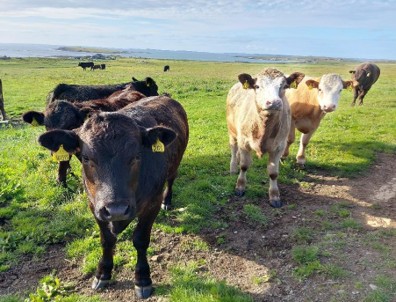
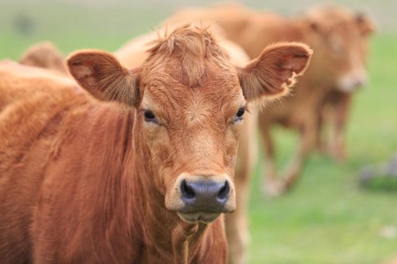
Interesting cases in December

Cattle
Abortions/mastitis

  • There were five cases of bovine abortion attributed to Salmonella Dublin in December and Salmonella Mbandaka was isolated in one case.
  • Paired serology demonstrated seroconversion to Salmonella Dublin in two aborted Holstein dairy cows from a herd affected with an increased incidence of abortion and early embryo losses.
  • Trueperella pyogenes was isolated in cultures of foetal stomach contents of one of two calves aborted at approximately five months gestation. Both animals had previously had successful calvings in the herd. T.pyogenes is classed as an opportunistic pathogen which is a recognised cause of sporadic abortion in cattle. It can arrive haematogenously from a septic focus elsewhere in the body such as a liver or foot abscess. In a second submission T.pyogenes was isolated from foetal stomach contents and Coxiella burnetii (Q fever) was detected by PCR in foetal tissues. It was unclear if the positive PCR result was significant or an incidental finding. Histopathology is required for further investigation of suspected Q fever abortion cases.
  • Two cases of bovine abortion due to Bacillus licheniformis were diagnosed this month.
  • There was one case of bovine abortion due to Listeria monocytogenes. L.monocytogenes was isolated in a further abortion submission but in that case L.ivanovii was also isolated along with several other bacteria (Proteus sp, Aeromonas sp.) raising the suspicion of sample contamination in that case. To reduce contamination when collecting foetal stomach contents, it is advisable sample the contents using a sterile needle and vacutainer.
  • There were five cases in which foetal fluid tested positive for BVD virus in the antigen ELISA. In four of these cases a bacterial cause of abortion was identified in cultures Salmonella Dublin (2 cases), Bacillus licheniformis (one case) and Listeria monocytogenes (one case). The connection between the foetal BVD results and the bacterial cultures is not completely clear, though it was considered that a transient BVD viraemia could cause immunosuppression and potentially increase the risk of bacteria gaining entry to the uterine environment.
  • Antibodies to Neospora caninum were detected in foetal fluid in two cases indicating in-utero exposure of the foetus to this pathogen. Neospora caninum was detected by PCR in foetal tissues in at least one submission this month. Confirmation of Neospora abortion can be achieved through histopathology on fixed tissue.
  • There was a significant rise in antibodies to IBR on paired serology in the IBRgE test in an aborted dairy cow which went from a moderate positive to a high positive result indicating likely recent exposure to wild type IBR. IBR virus can cause placentitis and infect the foetus leading to abortions. IBR can also cause abortions secondary to pyrexia/illness without IBR infection of the foetus. In the case of foetal infection with IBR this can be confirmed by histopathological examination, immunohistochemistry of placenta and/or foetal tissues and PCR testing.
  • Pasteurella multocida was isolated in cultures of mastitic milk. Pasteurella multocida is a rare cause of mastitis in cows. As it can arise from teat injuries, prevention of teat damage is the most effective method of control of this pathogen. Cow-to-cow spread can occur, and so affected animals should be milked last or with a separate unit. Severe cases can result in endotoxaemia and can be fatal.
  • Klebsiella pneumoniae was isolated from two out of four milk samples from dairy cows in a herd which was experiencing a severe outbreak of mastitis. Klebsiella pneumoniae is considered a major pathogen in bovine mastitis and can cause severe clinical disease. It can cause long-term drop in milk production through chronic disease. Udder hygiene and maintaining a clean, dry environment are important measures for control of Klebsiella mastitis.

Respiratory disease

  • A postmortem tracheal swab was received from a British blue store calf which had been treated for signs of pneumonia but later died. A necrotic tracheitis identified at gross postmortem was suggestive of a diagnosis of IBR and this was confirmed by PCR testing of a tracheal swab. IBR was detected in fourteen further submissions in December.
  • RSV was detected by PCR in over fifty submissions this month. Tissues were received from an approximately six-month-old, pre-weaned, Limousin suckler calf, which was the first death to occur in the group which were showing signs of respiratory disease. The calves had been housed two weeks earlier and had received an intranasal vaccine at that time. Postmortem examination of the case showed marked cranioventral consolidation and hyperinflation and emphysematous bullae in the caudal lungs. PCR testing of lung tissue detected RSV along with Mycoplasma bovis, Histophilus somni and Pasteurella multocida. Histophilus somniwas isolated in cultures of lung tissue. Histopathology of lung identified changes typical of severe bacterial bronchopneumonia consistent with that caused by Histophilus somni and Pasteurella multocida identified on culture /PCR testing. There was mild airway necrosis which could be due to RSV infection, but it was noted that the changes could also be caused by bacterial pathogens. Immunohistochemistry was suggested to further investigate of the role of RSV in this death.
  • PI3 was detected in over twenty submissions this month either as the sole viral pathogen or in some cases in combination with Coronavirus or RSV.
  • Severe bacterial bronchopneumonia was identified as the likely cause of death of a three-day-old calf. Abundant gram-negative coccobacilli were present and possible pathogens included E.coli, a member of the pasteurellaceae, or salmonellosis. Bacteriology of fresh tissue was suggested to further investigate. There was evidence of meconium in the airways suggestive of distress at calving. Inhalation of meconium, lack of aeration of the lungs and associated inflammation can lead to weakened lung function and increased susceptibility to bacterial infection.
  • Postmortem tissues were received from a fifteen-month-old Holstein dairy heifer. Severe consolidation of the lungs and a fibrinous exudate were observed at postmortem examination. Mannheimia haemoltytica was detected by PCR testing. Histopathology of lung tissue identified a severe fulminating bacterial pneumonia consistent with the Mannheimia haemolytica detected on PCR (Figure 1).

Section of lung from a heifer

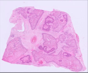
Figure 1: Section of lung from a heifer showing extensive inflammation and necrosis typical of Mannheimia haemolytica fulminating pneumonia.

  • Lungworm larvae with appearance consistent with Dictyocaulus viviparus were observed by microscopy in faecal samples from suckler cows which had a previous history of clinical disease due to lungworm and were noted to be coughing again at housing. D.viviparus larvae were detected by PCR in further two cases and a further three submissions identified animals that were seropositive for antibody to D.viviparus , consistent with lungworm exposure.

Gastrointestinal disease

  • Parasitic gastroenteritis was thought to be the cause of low body condition score and ill-thrift in a single animal in a group of 18-24-month-old cattle on an organic unit. The Strongyle egg count in a faecal sample was 1800epg, consistent with a heavy gastrointestinal parasite burden. There was evidence of hypoalbuminaemia on biochemistry which was likely to be linked to the gastrointestinal parasitism. In addition to the parasitism, the animal had evidence of low copper and selenium status and a pooled sample for inorganic iodine from the cohorts indicated that there was a low iodine intake in the group.
  • Salmonella 61:k:1,5,7 was isolated from a faecal sample from a yearling Limousin cross steer with a three-week history of weight loss and scour. A potentially significant Strongyle egg count of 250epg had been recorded at the practice. This serotype of Salmonella is considered host adapted to sheep and is very uncommonly reported in cattle. It was queried whether the animal was co-grazing or had any other contact with sheep.
  • In one submission Salmonella Typhimurium was isolated from faecal samples. Six cows on the farm had presented with acute onset scour and profound milk drop within two days.
  • A pooled faecal sample from three calves tested positive for Cryptosporidium parvum, Rotavirus and Coronavirus antigens.
  • Bovine coronavirus was detected by PCR in faecal samples from scouring freshly calved heifers, confirming a suspected diagnosis of Winter dysentery.

Skin and eye disease

  • Cutaneous lymphoma was diagnosed in a mature dairy cow with skin lumps. Sporadic lymphoma/leukosis usually affects animals younger than 4 years of age, with multicentric, thymic and cutaneous forms less commonly seen. The practitioner was advised to contact their local APHA office in case further investigation regarding the (very unlikely) possibility of the notifiable disease enzootic bone leukosis (EBL) was required.
  • Tissues were received from a ten-year-old Highland cow with a friable, necrotic mass cranial to the right horn base. The mass had previously been excised but had regrown to about four time its previous size to approximately 10cm diameter. It was noted that the mass had a hard overlying scab but the underlying tissue was soft and haemorrhagic. Histopathology of a section of the tissue identified the mass as a likely squamous cell carcinoma, though the small sample prevented definitive diagnosis in this case. SCC in cattle are usually locally invasive but don’t tend to metastasise until later in the course of disease. Spread in the first instance is usually to local lymph nodes.
  • Dermatophyte sp. was detected by PCR of hair plucks/skin scabs from a twenty-month-old bull with round, raised scaly skin lesions suspicious of ringworm infection. Mild pruritis was also observed in this case.

Systemic and miscellaneous disease

  • Mannheimia haemolytica was isolated in cultures of lung tissue from a weaned calf which was noted to have been a poor doer for some time prior to death. The isolate was thought to be a significant finding as a possible cause of pericarditis (and possibly peritonitis) which were seen at gross postmortem examination.
  • Salmonella Dublin was isolated from a liver swab from an eight-week-old dairy cross calf. The liver had appeared grossly enlarged and the gall bladder distended at postmortem examination.
  • Blackleg caused by Clostridium chauvoei was diagnosed by FAT (Fluorescent antibody testing) of muscle tissue sampled from a cow which was one of four from the same holding to have ‘dropped dead’ with no premonitory signs within twenty-four hours.
  • A yearling Limousin cross animal showed signs of bilateral blindness with corneal opacity, pyrexia (>40.50C), crusting of the muzzle, drooling and condition loss over a period of more than ten days. Testing confirmed that the animal had seroconverted to OvHV-2, making Malignant Catarrhal Fever (MCF) highly likely to be the cause of these clinical sings. MCF was diagnosed by PCR testing in four cases this month.
  • A round cell tumour most likely to be lymphoma was identified on histopathology testing of tissues from a five-year-old dairy cow. There was a history of rapid condition loss, pyrexia and lymph node enlargement of the prescapular, cervical and mandibular lymph nodes. Cardiomegaly and endocarditis were seen on postmortem examination and the lung tissue appeared pale. As with the cutaneous lymphoma case above, reporting to APHA was recommended in case further investigation was required to rule out enzootic bovine leukosis.

Sheep and goats

Reproductive disease, mastitis

  • Listeria monocytogenes was detected by PCR in tissues from a pygmy goat foetus that was aborted around six weeks prior to the kidding date. L. monocytogenes abortion is typically associated with spoiled or soil contaminated silage or grazing on muddy pasture.

Respiratory disease

  • Post-mortem tissues were received from a nine-month-old Mule ewe lamb after multiple deaths had occurred in a flock with a history of Haemonchus infestation, sheep scab and suspected clostridial enterotoxaemia. This increased mortality had occurred two to three days after dipping. On PME, the lungs were consolidated and abscessed. Histopathology revealed extensive suppurative and plasmalymphocytic bronchointerstitial pneumonia with bronchiolitis fibrosa obliterans, changes most associated with atypical/enzootic pneumonia. This has a multifactorial aetiology including Pasteurella multocida, Mannheimia haemolytica, Bibersteinia trehalosi, staphylococci, streptococci and Mycoplasma ovipneumoniae. Clinical consequences vary from reduced live weight gain and a mild cough to severe pneumonia and death which is usually associated with secondary bacterial infection. Recent herding and introduction of new animals into the flock can be associated with outbreaks. The bronchiolitis fibrosa obliterans indicated historic severe necrotising bronchiolitis which in sheep is most often caused by a virulent viral infection such as RSV or PI3.
  • OPA was confirmed on histopathology in a four-year-old ewe with suspicious signs on PME. The causative retrovirus is thought to be transmitted mainly by the respiratory route, probably via aerosol droplets and infection enters the flock through the introduction of sub clinically affected animals. On first introduction to a naive flock, between 30-80% of animals may succumb. However, once endemic, ongoing losses typically vary between 1 and 5% although they can reach as much as 20%. The virus does not survive for long in the environment and so depopulation of in-contact animals and repopulation from known disease-free flocks is the most successful method of control, here economically feasible.
  • A six-month-old Castlemilk Moorit lamb died after a 24-hour period of lethargy and slightly increased respiratory effort. Histopathology of lung detected multifocal acute random (embolic) pneumonia and hepatitis with intralesional gram-negative bacteria suggestive of septicaemic pasteurellosis due to Bibersteinia trehalosi. Potential stressors precipitating disease include handling, transport, mixing, movement to good grazing, and a change to cold, wet weather.

Gastrointestinal disease

  • Parasitic gastroenteritis was reported in sheep flocks and goat herds this month. In one Boer goat herd, an ill thrifty doe with pale mucous membranes that had recently been treated with Zolvix had a strongyle egg count of 4150 epg, raising the suspicion of haemonchosis and also of possible treatment failure. Haemonchus eggs were detected in a faeces sample from a six-month-old lamb that died suddenly with generalised pallor of the carcass found on PME; of a strongyle egg count of 5488 epg, 29% were determined to be Haemonchus eggs by the ovacyte method.
  • Fasciolosis was reported in several sheep flocks on faecal microscopy and coproantigen ELISA testing.
  • A total of 58 submissions for Johne’s disease serology and faecal PCR testing from sheep and goats were received, with 13 submissions, including a total of 22 individual samples, testing positive. In one case, a male pygmy goat was scouring for a little over a week and was very dehydrated. There was no improvement with potentiated sulphonamides, NSAIDs and oral rehydration and the animal became hypothermic, so the goat was euthanased. The animal was positive for Johne’s on serology and faecal PCR. In a second pygmy goat herd, after finding two positive animals, the healthy breeding stock was screened and 8 out of 22 animals (35%) were seropositive. In another case, a sheep flock of 150 – ewes and ewe lambs were in poor condition and there had been several deaths over a four-to-six-week period. There had also been poor fertility over the last two to three years. Johne’s disease was suspected and a pooled faeces sample from nine animals was positive on Johne’s PCR.

Skin and eye disease

  • A group of 12 ewes and four ewe lambs developed keratoconjunctivitis. The sheep had been treated with long acting oxytetraycline and the condition improved but since returned. Subsequent treatment with tulathromycin had minimal response. Mycoplasma conjunctivae was detected by PCR on submitted ocular swabs. M. conjunctivae typically enters a flock via healthy carriers and is rapidly transmitted between animals by direct contact and potentially vectors such as flies, pollen and dust. Recurrent infections are common as, despite apparent resolution following treatment with e.g. oxytetraycline, infection may persist within the conjunctival sac for several months.
  • A ram lamb in a commercial flock had multiple raised, proliferating growths on all four limbs with the most extensive lesions being below the hocks on the hind limbs. Histopathology detected severe ulcerative, hyperplastic, hyperkeratotic and crusting dermatitis with ballooning degeneration and intralesional eosinophilic intracytoplasmic inclusion bodies consistent with orf. All lambs were reported to have been orf vaccinated in the first week of life.
  • At least two cases of sheep scab were confirmed on skin microscopy. Several sheep were also seropositive for sheep scab with reported clinical signs including pruritus, scab formation, wool loss, behavioural changes and condition loss.
  • Two cases of periorbital eczema due to Staphylococcus aureus were reported in sheep flocks. This is usually secondary to trauma e.g. bumping heads from pushing to get into feed troughs leading to skin abrasions and allowing entry of bacteria. In one flock, erosive lesions were also seen on the ears as well as around the eyes.

Periorbital eczema in a ewe

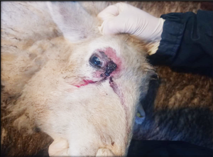
Periorbital eczema in a ewe (Photo by Laura Barlow, Castle Farm Vets, Barnard Castle)

Systemic and miscellaneous disease

  • Cerebrocortical necrotising encephalopathy was diagnosed on histopathology of brain from one of several lambs dying in a flock. The most common cause of this is cerebral cortical necrosis (CCN) due to thiamine deficiency associated with dietary changes.
  • Seroconversion to Maedi visna was reported in several flocks. In many cases, positive results were in flocks where MV was already known to be present. However, evidence of exposure to MV was found for the first time in a flock where there were reported to be higher numbers of thin ewes.
  • Seroconversion to CLA was reported in at least two sheep flocks and two goat herds. In all four cases, an animal with a lump/abscess in the parotid region was reported.
  • After 12 eight- to nine-month-old lambs died over two days, blood samples were received from one lamb in the flock. The lamb was seropositive for louping ill virus, with the majority of antibody present being IgM, consistent with recent exposure and infection. The lamb also had a very low vitamin B12 level consistent with cobalt deficiency.
  • Post-mortem tissues were received from an 11-year-old, male adult pygmy goat which was euthanased after a 24-hour illness culminating in collapse, dyspnea, groaning and injected red conjunctivae. On farm PME detected small cream homogenous nodules throughout the liver and spleen and, on histopathology, the parenchyma of both organs was effaced by a densely cellular, unencapsulated, infiltrative proliferation of neoplastic epithelial cells, consistent with an epithelial tumour, possibly a cholangiocarcinoma (Figure 2&3).

Low power view of the spleen

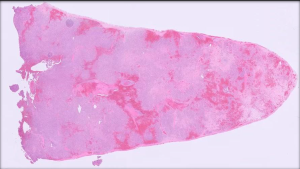
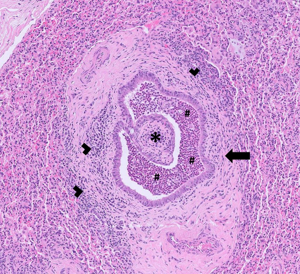
Figure 2: Low power view of the spleen showing near diffuse effacement of normal architecture by a proliferation of neoplastic epithelial cells.Higher power view of the neoplastic cells

  • Uroliths predominantly of calcium carbonate were surgically removed from the urethra of a pygmy goat undergoing perineal urethrostomy. Predisposing factors to these uroliths include castration in males and legumes in grazing or forage.

Camelids

  • Fasciolosis was detected in a thin, recumbent adult female alpaca on faecal coproantigen testing. It was also detected in another herd on faecal microscopy.
  • Post-mortem tissues were received from a 16-month-old female alpaca with suspected lymphosarcoma. Histopathology detected severe granulomatous enteritis, lymphadenitis and lymphangitis with abundant acid-fast bacteria, most consistent with Johne’s disease.(Figure 4). A second case was confirmed in an alpaca on faecal PCR.

Abundant acid-fast bacteria

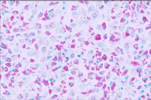
Figure 4: Abundant acid-fast bacteria stained pink with Ziehl-Neelsen special stain, in the intestine of an alpaca with Johne’s disease

  • Six out of seven faeces samples from Bactrian camels in a zoological collection had positive strongyle egg counts of which were five were well over 1000 epg two weeks after anthelmintic usage, consistent with treatment failure and also raising the possibility of haemonchosis. A similar case was seen in a camel in a private collection which had a strongyle egg count of 4750 epg ten days after anthelmintic treatment.

Other Species

  • An inappetant eight-year-old sow had an Ascaris suum count of 5550 epg. This is a very high ascarid egg count and occasionally large, adult worms can form a mass blocking the intestinal lumen.
  • MCF (OvHV-2) was diagnosed in a reindeer on PCR testing. No history was provided for the case, but reindeer are known to be susceptible to OvHV-2 infection and cases are invariably fatal.
  • Postmortem samples were received from a three-month-old Axis deer after four calves from two deer species at a zoological collection were euthanased due to weakness and collapse. PME had detected pale skeletal muscles and renal cortices, and histopathology was consistent with a moderately severe tubulointerstitial nephritis. Potential causes included acorn poisoning and haematogenous spread of bacterial infection.
  • Post-mortem tissues received from a three-week-old hand-reared female Elds deer calf in a zoological collection. The calf had suffered acute onset lethargy and neurological signs and was euthanased. Although no scour was noted, PME revealed haemorrhagic enterocolitis. A mild suppurative splenitis and colitis, and mild, subacute interstitial pneumonia were found on histopathology consistent with salmonellosis, with isolation of Salmonella Coeln from the intestine. This is a relatively rare serotype of Salmonella that has been found in cattle, horses and primarily poultry.
  • Post-mortem tissues were received from a female rabbit. The lower incisors had been extracted and the rabbit subsequently developed osteomyelitis. On PME, there was evidence of pulmonary abscessation and pleural empyema, with histopathology detecting extensive, severe, subacute necrosuppurative pneumonia with intralesional gram-negative coccobacilli, consistent with the isolation of Prevotella heparinolytica on culture. P. heparinolytica has been associated with mandibular and maxillary abscesses in rabbits and may have seeded in the respiratory tract following haematogenous spread or inhalation.

Deficiencies and toxicities

  • Samples were received from cows in a suckler herd which had issues with weak calves and previously diagnosed copper deficiency. Despite administration of copper containing boluses and free access to lick buckets over the summer, three of the five animals tested had significantly low copper levels.
  • Hypocupraemia was identified in an eight-year-old male reindeer which was scouring and losing weight. There was no evidence of gastrointestinal parasitism in worm egg counts carried out in the practice.
  • Low GSH-Px indicative of selenium deficiency was seen in a six-month-old male cross Hereford calf noted to be inactive and slow. A similar calf tested earlier in the year also had low GSH-Px. Screening of cows found four out of five had low GSH-Px levels (the remaining animal had a GSH-Px level of 31U/ml RBC, just above the cut off value for deficiency of 30U/ml RBC). There was no evidence that the animals were copper deficient.
  • Low pooled PII, suggestive of likely iodine deficiency was recorded in a group of cows from which several ‘slow and dopey’ calves had been born, and a single stillbirth had occurred. PII was also low on a second farm where 20 out of 50 cows were not in calf.
  • Reports of Acorn toxicity cases continued into December. One submission was from a farm on which a group of store cattle had broken out their field and escaped into an oak wood. Eight animals died and a further five showed signs of ongoing malaise. Blood testing indicated that all five had moderate to severe azotaemia indicative of impaired renal function and guarded to poor prognosis for recovery.

Follow us on Facebook

https://www.facebook.com/Axiomfarmvetlab/

This newsletter is sent by e-mail to each vet practice but if you would like a copy sent to your individual e-mail account please contact us at dsfarm@axiomvetlab.co.uk and we can add you to our circulation list.

Related Posts