Farm April Newsletter 2026

Dedicated farm line – 01626 357776

Follow us on Facebook

https://www.facebook.com/Axiomfarmvetlab/

This newsletter is sent by e-mail to each vet practice but if you would like a copy sent to your individual e-mail account please contact us at dsfarm@axiomvetlab.co.uk and we can add you to our circulation list.

Would you like to send the Axiom farm vets photos of PMEs to allow us to assist with sample selection and diagnosis? Please send photos by WhatsApp to 07944 649222.

You may find this is a more efficient way of making requests than phoning the farm team, saving you time in your busy day. Our farm team also find it a more efficient way of dealing with your requests.
The email address for test requests is: dsfarm@axiomvetlab.co.uk 

Axiom Farm Vets

If you wish to speak to a particular farm vet about one of your cases, you can consult the table below to see when our vets are usually available. A duty farm veterinary advisor is always around on a Saturday morning from 9am-12pm.

Farm Vets

Feedback on our services

As part of our continuing effort to help improve our services to clients within the Laboratory Division we have put together a short survey which will be sitting live permanently for anyone to complete whenever they have a few minutes spare. This can be filled in by anyone who uses any of our services and we encourage feedback to
help us understand what we are doing well and where we need to make improvements. The QR code and link to the survey will remain live as a continuous tool to enable us to always gather feedback. If anyone has any questions about this, they are welcome to contact either of the Quality Managers at the Laboratories, Claire
Richardson for Axiom Veterinary Laboratories and Susan Reeve for Finn Pathologists. Thank you in advance for helping us to improve our services.
https://www.surveymonkey.com/r/Laboratory_Satisfaction_Survey

Feedback QR Code

BTV testing guidance

We wish to remind vets that as a commercial laboratory we are unable to offer any Bluetongue testing either on blood samples or abortion material. If we receive samples from cases in which Bluetongue is suspected, we are required to safely dispose of the samples and cannot provide any further testing. We are always happy to discuss cases over the phone before submission of samples. Clinical suspicion of BTV infection must be reported to APHA. Further information on investigating poor reproductive performance in cattle and sheep during Bluetongue outbreaks can be found at the following link:

BTV Web Link

Foetal stomach contents

To maximise the chance of a diagnosis in abortion cases, foetal stomach content (FSC) is one of the most useful samples we can receive. It is used primarily to identify bacteriological and fungal causes of abortion. It is an important component of our abortion PCR package (Test code: FABORT) where it is used in a ‘pool’ along with fresh foetal brain and liver tissue. In a freshly aborted foetus, the abomasal contents will be relatively protected from environmental contamination as opposed to, for example, placental tissue which tends to be heavily contaminated. When sampling FSC the risk of contamination can be minimised by using a sterile vacutainer and needle punctured though the stomach wall to sample the fluid inside. Where insufficient fluid is present then an alternative is to nick the stomach wall with a clean scalpel and introduce a swab (charcoal or other swab with transport media suitable for bacteriology) through the wall to sample the contents. FSC is only suitable for testing in abortion cases and cases at term where the animal was known to have been stillborn. Stomach contents from animals that have been alive for several hours/ days are not suitable for testing as they may have received milk/milk replacers or attempted to suckle contaminating of the stomach contents and leading to less than useful results e.g detection of the agent in milk/colostrum.

Molecular mastitis test

Unfortunately, the Mastitis 16 pathogen PCR referral test is no longer available to us. As an alternative you may wish to consider the Mastitis Rapid Molecular test which is available at Axiom (Test code: FAMAS). This test detects the presence of six key mastitis pathogens, and the turnaround time is no longer than the following working day. This can allow a targeted course of treatment based upon the pathogens present, aiding responsible use of antibiotics and resulting in less discarded milk. As well as potentially saving money there are benefits for herd health in the long term. The agents detected are E. coli, Staphylococcus aureus, Streptococcus uberis, Streptococcus dysgalactiae, Streptococcus agalactiae and Klebsiella pneumoniae. The cost of the test is £45 & VAT (test code FAMAS).

Interesting cases in February

Cattle
Abortions/mastitis

  • Necrotising encephalitis and hepatic necrosis were observed on histopathology of brain and liver tissue of an aborted calf foetus. The 500-cow dairy farm from which the foetus was submitted had recorded five abortions in late 2025. At that time samples from two aborted calves did not identify any causal pathogen. A further four abortions occurred over ten days in February. Changes on histopathology were suggestive of ongoing hypoxia of the foetus, most likely caused by placental insufficiency. Placental insufficiency is the result of inadequate perfusion of the placenta, leading to suboptimal oxygen and nutrient delivery to the foetus. Possible causes could include nutritional stress or deficiencies, infectious agents (no inflammation seen and no evidence of bacterial or fungal organisms with special stains), toxicoses, or other insults.
  • Salmonella Anatum was isolated from one case of bovine abortion this month. This serotype is usually associated with contaminated feedstuffs. Wild birds and badgers have both been implicated in the carriage of this serotype.
  • Actinobacillus seminis was isolated from foetal stomach contents in one case of bovine abortion. Actinobacillus seminis is reported as a cause of epididymitis in sheep and has also been associated with bovine abortion and was considered a relevant finding in this case.

Respiratory disease

  • An adult dairy cow presented in late gestation with a clear nasal discharge, pyrexia of 410C and was anorectic. Approximately six cows and one bull were similarly affected in the herd of around 80 animals. IBR infection was suspected and confirmed by PCR testing of a nasal swab from the sick cow.
  • Postmortem tissues were submitted from two one-month old Holstein- Friesian bull calves which were seen with signs of respiratory distress in the evening but died overnight despite treatment for pneumonia. The submitting vet noted that there was a suspicion that both calves had suffered from failure of colostral absorption. Mannheimia haemolytica and Pasteurellla multocida were detected by PCR in lung tissue of both calves. Histopathology identified a severe fulminating bacterial pneumonia consistent with the PCR results.
  • More than fifteen cows in a 500-cow dairy herd were affected with respiratory symptoms which had started one week before. Samples were submitted from the first cow to die in the outbreak. Mannheimia haemolytica was detected in lung tissue by PCR testing and the finding was of likely significance in the outbreak.
  • Respiratory signs were seen in a group of approximately twenty suckler calves running in a cow and calf group. A few of the calves had already been treated for presumed pneumonia by the owner. Rectal temperatures were taken from all the calves on the day of examination and the majority had pyrexia over 39.20C. Untreated calves with temperatures >400C were selected for sampling and a pool of four swabs tested in the respiratory pathogen PCR package was positive for RSV, indicating that this pathogen was likely responsible for the respiratory disease outbreak.
  • Postmortem tissues were received from a post-weaned suckler calf from a group in which respiratory signs had been seen. This was the second calf to die within a 48-hour period. RSV along with Pasteurella multocida and Mannheimia haemolytica were detected by PCR of a plain lung swab. Marked, extensive, subacute, necrotising and suppurative bronchointerstitial pneumonia with interstitial emphysema was seen on histopathology. These changes supported a diagnosis of RSV infection and a secondary bacterial component, consistent with the PCR results.
  • RSV was detected by PCR testing of lung tissue from a seven-month-old limousin cross suckler calf. Consolidation of the lung had been observed at postmortem examination. Histopathologically, airways contained small numbers of neutrophils with admixed cell debris. Airway epithelium had been lost and replaced by fibrin. Alveoli were variably collapsed, diffusely congested, and contained rich proteinaceous fibrin material with few red blood cells and scattered macrophages and neutrophils. Alveolar septa were extensively necrotic, replaced or lined by thick layers of fibrin (hyaline membranes). Multifocal Type II pneumocyte hyperplasia was present. Small numbers of mixed inflammatory cells surrounded blood vessels. Interlobular septa were markedly expanded by dilated lymphatics, clear space and eosinophilic proteinaceous fluid. Overall, these changes were considered severe and typical of RSV infection as per the PCR result.
  • Approximately ten of a group of forty British Blue cross heifer calves were affected by severe pneumonia which showed little improvement with tulathromycin treatment. Lung tissue from a calf which died was tested by PCR for respiratory pathogens and was positive for Mycoplasmopsis (formerly Mycoplasma) bovis, Mannheimia haemolytica and Pasteurella multocida. Lung tissue was evaluated by histopathology. Changes to the lung were severe and strongly suggestive of virulent Mycoplasmopsis bovis infection with extensive eosinophilic coagula occluding airways and associated plasmalymphocytic peri-airway infiltrate (Figure 1). Considering the PCR results, it was suggested that this case could be a combined Mannheimia haemolytica and Mycoplasmopsis bovis infection. It was noted that Mannheimia haemolytica infection can worsen the pneumonia caused by M.bovis.

Lung tissue is near completely effaced

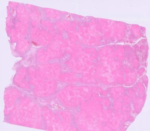
Figure 1: Lung tissue is near completely effaced by bright pink eosinophilic coagula (caseonecrotic material) in a calf with virulent Mycoplasmopsis bovis pneumonia.

Gastrointestinal disease

  • Post-mortem tissues were received from a two-week-old, Holstein steer calf. It was one of 120 calves which had arrived one week previously at the calf rearing unit. Calves were divided into four groups with automatic feeders in straw pens. There was access to eight litres of milk per day along with calf grower pellets. There has been a recent rise in the incidence of bloat, with at least ten calves affected and this animal being the fourth to die. At postmortem examination the abomasal mucosa appeared inflamed but no obvious haemorrhage or ulceration was seen. Histopathology identified a moderate, extensive, suppurative abomasitis with submucosal emphysema and intralesional bacterial with appearance typical of Sarcina sp. Sarcina bacteria are often found associated with abomasal bloat and abomasitis in young calves, and the aetiology is still not fully understood. Several predisposing factors are thought to be involved including compromised immunity from failure of passive transfer, bacterial infection of the abomasal wall, and vitamin and mineral deficiencies. The role of bacteria, such as Clostridium perfringens, Clostridium fallax, Clostridium sordellii and Sarcinia spp., in these cases is unclear, but mixed infections have been associated with disease. Entry and proliferation of the bacteria are likely associated with damage to the abomasal mucosa. In artificial milk fed calves this is most often caused by poor milk hygiene, poor quality milk, large volumes of milk fed or milk fed at the wrong temperature.
  • Postmortem tissues were received from a 7-10 day old calf which had died suddenly. An abomasal ulcer was found on post mortem examination, and fungal hyphae were found within the associated inflammation on histopathology (Figure 2). Abomasal ulceration in young dairy calves are thought to develop secondary to stress, poor milk feeding practices, or because of coarse feed or grain overload.

Fungal hyphae found in the wall of the abomasum

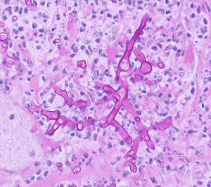
Figure 2 : Fungal hyphae found in the wall of the abomasum of a calf with abomasal ulceration (PAS stain)

  • Enterotoxigenic Escherichia coli (ETEC) infection was diagnosed by detection of E.coli F5 (K99) antigen in faecal samples from two diarrhoeic calves from the same farm. Several calves had been affected by scour with deaths occurring at 24-48hours of life. E.coli was cultured from the samples and in both cases the isolates showed evidence of invitro resistance to multiple antibiotics.
  • An untypable Salmonella species with the antigenic formula 6,7,:z:10:- was isolated from faeces of an adult dairy cow showing signs of acute milk drop and green water scour. Twenty-five of 350 cows were reported to be showing similar signs. Occasionally Salmonella isolates can be difficult to serotype and this may occur if the isolate is unusual, damaged or doesn’t behave in the expected manner under test conditions. Further testing was advised if the problem was ongoing or recurred.

Skin and eye disease

  • A pedunculated mass which had appeared over an estimated two weeks on the prepuce of a two-year old Aberdeen Angus bull, was excised and sent for histopathological examination. The mass was identified as a fibropapilloma. (Figure 3) Fibropapillomas are hyperplastic lesions caused by papillomavirus infection. Penile fibropapillomas in bulls are most often diagnosed at one to two years of age. They can spontaneously resolve, but this occurs after a variable time and recurrence can be a problem.

A cross-section of a penile fibropapilloma

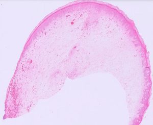
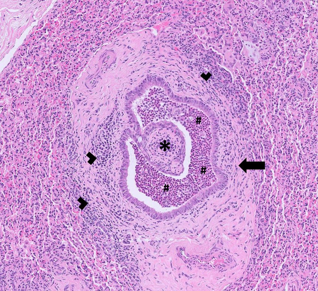
Figure 3: A cross-section of a penile fibropapilloma from a bull showing a proliferation of loosely packed mesenchymal cells, covered by a hyperplastic epithelium.

  • Soft tissue sarcoma of the penis was identified on examination of tissues received from a twelve-month-old crossbreed finishing bullock. The submitting vet noted that there appeared to be multiple masses present which were dark red in colour and inflamed. Histopathology confirmed the mass as a spindle cell tumour and nuclear atypia, karyomegaly, necrosis, and frequent bizarre mitoses suggested malignancy. The most likely diagnosis was fibrosarcoma, but other soft tissue tumours were also possible (e.g. peripheral nerve sheath tumour, haemangiopericytoma, myxosarcoma, leiomyoma/-sarcoma, undifferentiated sarcomas). Cases of penile fibrosarcoma are rare but have been previously reported in cattle. Treatment involves prompt, complete surgical removal, but these tumours have a high rate of recurrence due to their aggressive, infiltrative nature.

Systemic and miscellaneous disease

  • One of two Aberdeen angus cross calves which were bought in at two weeks of age and had been on farm a further fortnight tested positive on serum in the BVD antigen ELISA indicating BVD viraemia. The result was a low positive with an optical density of 0.9 OD. (Positive: >0.3OD). In this test positive results close to the positive/negative cut off are more likely to be consistent with transient infections. However, persistent infection cannot be ruled out as high levels of maternally derived antibody could potential mask viraemia to some degree. A second BVD antigen test must be carried out after at least three weeks and if antigen positive at that time BVD PI status of the animal is confirmed. On this farm it was observed that two or three from each group of bought in calves would show signs of illness and there was a higher than target mortality. It was unknown how much BVD could be contributing to the problem in this case as there was a complex history of issues with calfhood illnesses including cryptosporidium and coccidiosis.
  • Malignant catarrhal fever (MCF) was confirmed by PCR testing of a blood sample from a six-month-old Jersey bull. The animal showed signs of acute pyrexia, bilateral corneal oedema and copious fetid nasal discharge, all typical clinical signs of MCF due to OvHV-2 infection in cattle.

Sheep and goats

Abortions, reproductive disease, mastitis

  • Multiple cases of enzootic abortion were diagnosed this month, primarily on examination of MZN-stained smears of placentae but also on PCR testing, including some vaccinated ewes. Vaccination will tend to reduce the number of abortions that occur but will not eliminate infection from sheep that have become infected the previous year.
  • Multiple cases of abortion associated with Campylobacter fetus were also diagnosed this month on culture and on PCR testing.
  • At least three cases of abortion due to E. coli were reported. Typically, this is associated with sporadic abortions following ascending infection and opportunistic invasion of the gravid uterus. An Acinetobacter sp. in one flock and a Bacillus sp. in a second flock were also potential causes of sporadic abortion following opportunistic ascending infection after being cultured in pure growth from foetal stomach contents.
  • At least two cases of Toxoplasma gondii abortion were diagnosed, one on foetal fluid serology and one on PCR of placenta.
  • Abortions associated with the Listeria species, L. monocytogenes and L. ivanovii were diagnosed this month after isolation from foetal stomach contents. These are typically associated with the feeding of spoiled or soil contaminated silage or feeding on muddy ground.
  • High rates of seropositivity for Border disease were reported in ewes in flocks with poor scanning results and the birth of non-viable lambs.
  • Seropositive ewes for Tick Borne Fever were reported in a Lleyn flock and a Welsh Mountain flock with high barren rates.
  • Corynebacterium pseudotuberculosis, the causative agent of CLA, was detected in a milk sample from a Dutch Spotted ewe. This may occur when an abscess present in the mammary gland ruptures.

Respiratory disease

  • Five deaths occurred in a flock of 100 ewes over the course of a week. Affected animals presented with blindness, staggering, lethargy and sometimes excessive salivation. Histopathology of lung submitted from one ewe was consistent with ovine pulmonary adenocarcinoma with secondary fibrinosuppurative necrotising bronchopneumonia.
  • Muellerius capillaris were detected on Baermann’s testing in at least two goat herds, one of which reported coughing. M. capillaris tends to be more pathogenic in goats than sheep and heavy burdens may cause respiratory signs.


Gastrointestinal disease

  • Atrophic enteritis associated with the ileum and ileocaecocolic junction in samples received from lambs was diagnosed on histopathology in an outbreak of neonatal scours in a Suffolk flock. The cause was not apparent from the sections, with no parasitic structures observed, including coccidia and cryptosporidium. There were intraluminal bacteria, but these were nonadherent and non-invasive. Potential differentials included E. coli (although not directly observed), rotavirus and coronavirus. Rotavirus Type B had been detected in an earlier scour sample received from the flock and therefore was the likely cause.
  • Fasciolosis was reported in several flocks, diagnosed through both faecal microscopy and coproantigen ELISA. Where a history was provided, ill thrift was reported. In one sick, scouring Cheviot ewe there was also evidence of haemonchosis with 21% of a strongyle egg count of 1400 epg being confirmed as H. contortus eggs by the Ovacyte method.
  • Parasitic gastroenteritis was diagnosed in several flocks and herds this month. In one case, haemonchosis was confirmed in a ewe that had an in house WEC of 4400 epg and died despite treatment with closantel for suspected haemonchosis. 17% of the eggs were found to be consistent with Haemonchus on the Ovacyte method. Haemonchosis was also suspected in other sheep and goats with high strongyle egg counts but with no confirmatory testing carried out. Some sheep and goats with PGE also had high pepsinogen levels.
  • Johne’s disease was confirmed on intestinal histopathology in an Easycare ewe. A thickened ileum had been found on PME and histopathology detected a marked, chronic, diffuse granulomatous enteritis and lymphangitis with numerous acid-fast bacilli within macrophages within the small intestine, supporting a diagnosis of Mycobacterium avium subspecies paratuberculosis infection. Several more cases were diagnosed in sheep and goats on serology and faecal PCR.
  • Salmonella Typhimurium was isolated from the faeces of a ewe that died after developing acute diarrhoea after starting on silage.
  • Type D clostridial enterotoxaemia was suspected to be the cause of death in a three-year-old pygmy goat that died after a less than 24-hour period of haemorrhagic scour. Clostridium perfringens epsilon toxin was detected in intestinal contents. The goat was not vaccinated for clostridial disease.

Skin and eye disease

  • Several flocks had seropositive sheep on sheep scab serology, with signs including encrusting skin lesions, wool loss, pruritus and condition loss.
  • Bovicola chewing lice infestation was diagnosed on skin microscopy in a male pygmy goat with multifocal pruritic, scabbing skin lesions and in a herd of Golden Guernsey and pygmy goats that were pruritic despite treatment with ivermectin. Ivermectin is typically unreliable for treatment of chewing lice as the lice do not ingest blood. Synthetic pyrethroids would be expected to be more effective.
  • Staphylococcus aureus was isolated on culture of a swab from thickened skin in the groin of a pygmy goat and in a case of periorbital dermatitis in another pygmy goat that had been waxing and waning for nearly a year, responding to antibiotics but recurring in a few weeks.
  • Mycoplasma conjunctivae, the most common cause of infectious keratoconjunctivitis in sheep, was diagnosed on PCR of ocular swabs in four flocks. Two of the flocks reported recurrent infections despite systemic and, in one flock, topical oxytetracycline treatment. Cases can be difficult to successfully resolve due to failure to eliminate the bacterium from deep within the conjunctival sac.
  • Fine needle aspirates were taken from three of multiple skin lumps on a 12-year-old ram. Cytology detected marked expansion of intermediate to large lymphoid cells, confirming lymphoma in all three sites. Cutaneous extranodal lymphoma is rarely reported in sheep; when present it is typically part of multicentric disease. As seen in cattle, sporadic and enzootic forms of lymphoma may also occur in sheep and the submitting vet had reported a ‘farm history of lymphoma’. Although it could not be ruled out, involvement of Bovine Leukocytosis Virus (BLV) was considered unlikely, given the species and because the UK is considered clear of this virus.

Systemic and miscellaneous disease

  • In addition to the mastitis case reported above, CLA was detected in at least two further flocks and in an adult goat on serology and culture.
  • Seroconversion to Maedi visna virus was reported in several flocks. In one flock, three out of 24 sampled animals seroconverted to MV and three out of 24 seroconverted to Johne’s disease.
  • A 13-year-old wether had gradual onset ataxia over several days, occasionally falling over and circling but still able to walk and eat. On clinical examination, there was a delayed pupillary light reflex and an intact menace reflex. On haematology, there was a markedly elevated leucocyte count of 1127 x 109/l (RI: 5-11 x 109/l) of which the vast majority consisted of a large population of medium to large mononuclear cells with moderate amounts of basophilic cytoplasm and occasional indistinct nucleoli. This was strongly suggestive again of lymphoma.
  • Post-mortem tissues were received from a one-year-old, Texel cross ewe, one of 15 out of 900 ewes and ewe lambs to have died suddenly with no previous signs. PME detected consolidation in the lungs and histopathology had detected subacute interstitial pneumonia plus fibrinosuppurative and histiocytic colitis, both with intralesional bacteria. Listeria monocytogenes was isolated in mixed growth on culture of pleural fluid and the presence of short gram-positive rods in the intestinal wall and the lung supported a diagnosis of systemic listeriosis.
  • E. coli culture and sensitivity testing of cases of watery mouth yielded some multi-resistant isolates this month.

Camelids

  • Post-mortem tissues received from a female adult alpaca that was in very poor body condition, with liquid faeces and a grossly thickened large intestinal wall on PME. A strongyle egg count of 1100 epg was detected in the large intestinal contents and histopathology of the wall of the large intestine revealed an extensive fibrinosuppurative enteritis/colitis with crypt abscesses and crypt necrosis, and extensive mucosal fibrosis. These changes were severe, chronic and  ongoing and supported a diagnosis of parasitic gastroenteritis, with a likely secondary bacterial component. Further cases of parasitic gastroenteritis were diagnosed in alpacas on faecal egg counts.
  • Streptococcus infantarius was isolated from a swab taken from a chronic multi pocketed rump abscess in an alpaca that had failed to respond to potentiated amoxicillin. This has been reported as part of the gastrointestinal flora of alpacas but has also been reported as an opportunist pathogen in, for example, horses.

Other Species

  • Parasitic gastroenteritis was diagnosed on faecal egg counts in two reindeer. One of the two, an old female with soft faeces, had a strongyle egg count of 15,100 epg plus Capillaria and Trichuris egg counts of 250 and 50 epg respectively.
  • Ringworm was diagnosed on dermatophyte culture with the detection of Nanizzia nana (formerly Microsporum nanum) in an eight-year-old Tamworth sow with circular lesions on the shoulders, behind the left ear and the right hock, that had developed over a two-week period.

Deficiencies and toxicities

  • A South Devon cow which was around three weeks off calving presented with signs of loose dung, dullness and dehydration. Blood samples were taken for biochemistry which revealed mild azotaemia, a raised BHB of 8.04mmol/l (RI: 0.00-1.2mmol/l) and a raised NEFA level of 2792umol/l (RI: 0-600umol/l). The liver enzyme GLDH was also significantly raised at 456 U/L (RI: 0-25) indicating there was hepatocellular damage. The results were suggestive of pregnancy toxaemia in this cow with hepatic lipidosis being the possible cause of the raised GLDH.
  • Pregnancy toxaemia was confirmed by measurement of BHB in three ewes in late stages of pregnancy which had become weak, unable to stand and one died despite treatment with calcium and propylene glycol. All three ewes were reported to be in good body condition. Pregnancy toxaemia can occur in well fed over conditioned ewes in late pregnancy (body condition score of 4-5). Fat ewes have a decreased food intake in late pregnancy due to intraabdominal fat and the developing lambs leaving less space for the rumen. There is an increased risk of disease where foods with a high moisture content are being fed, such as silage or root crops. Concurrent hypocalcaemia is common.
  • Significant hypocalcaemia was diagnosed in two triplet bearing ewes. One of the two ewes was hypoalbuminaemic and the other had an albumin level at the lower end of the reference interval. Calcium levels were corrected to take low albumin into account. Using the calcium correction formula calcium levels were confirmed as low in both ewes.
  • In one dairy herd cows were reported to be showing signs of pica (abnormal/depraved eating behaviours). The causes of pica are not always clear, though phosphorus deficiency is commonly blamed. In this case three of twelve cows tested had evidence of hypophosphataemia and this was considered of possible significance. Sodium deficiency, low fibre intakes and mineral leaching due to heavy winter rain are also considered to be risk factors for pica in cattle.
  • Hypomagnesaemia was thought to be the cause of sudden death in a primiparous suckler cow out at grass with a nine-week-old calf at foot. The magnesium level in aqueous humour sample approximately two hours after death was 0.46mmol/l, suggestive that hypomagnesaemic tetany was the likely cause of death.
  • Several ten-month-old Romney store lambs were reported to be going off their legs and were in poorer than expected condition. An intention tremor was seen on attempts to rise and those that could rise had a stiff, stilted gait. The feet appeared to be good, and no swelling of the joints was seen and the lambs were reported to be otherwise bright. Four lambs were sampled and all had low GSH-Px levels, indicating hyposelenosis. Selenium deficiency was the likely cause of the musculoskeletal signs and possibly for the poor body condition. A pooled liver fluke serology sample from the lambs was positive for antibodies to Fasciola hepatica indicating exposure to liver fluke which could be a further contributing factor to poor condition.
  • One flock of Welsh mountain ewes had a disappointingly high barren rate of 12%, with twin pregnancies being reduced by two-thirds on previous years. Biochemistry testing revealed evidence of low selenium and low copper status in the flock as well as low albumins which could be the result of parasitic gastroenteritis, liver fluke or long-term low protein intakes. Three of five ewes sampled had raised pepsinogen levels consistent with some degree of abomasal damage due to parasitism. Pooled inorganic iodine testing indicated that the group had marginally low iodine levels in the diet.
  • Postmortem tissues were received from a bovine which was euthanised after showing neurological signs. There had been previous issues with ragwort toxicity on the farm and the suspicion of hepatopathy was supported at postmortem examination as the liver appeared rounded and had a ‘nutmeg’ appearance. Histopathology of liver tissue identified a chronic, severe, portal-bridging fibrosis with biliary hyperplasia, hepatocellular necrosis, nodular regeneration and megalocytosis consistent with a toxic insult and ragwort toxicity was considered most likely. Other toxicosis especially mycotoxicosis (e.g alfatoxicosis) would be a possible differential for these liver changes

Follow us on Facebook

https://www.facebook.com/Axiomfarmvetlab/

This newsletter is sent by e-mail to each vet practice but if you would like a copy sent to your individual e-mail account please contact us at dsfarm@axiomvetlab.co.uk and we can add you to our circulation list.

Related Posts