Farm June Newsletter 2026

Dedicated farm line – 01626 357776

Follow us on Facebook

https://www.facebook.com/Axiomfarmvetlab/

This newsletter is sent by e-mail to each vet practice but if you would like a copy sent to your individual e-mail account please contact us at dsfarm@axiomvetlab.co.uk and we can add you to our circulation list.

Would you like to send the Axiom farm vets photos of PMEs to allow us to assist with sample selection and diagnosis? Please send photos by WhatsApp to 07944 649222.

Note that we may wish to include photos sent to this number for the interesting cases section of this newsletter. If you do not wish your photo to be used, please let us know at the time of sending.

Email us with your additional test requests

You may find this is a more efficient way of making requests than phoning the farm team, saving you time in your busy day. Our farm team also find it a more efficient way of dealing with your requests.
The email address for test requests is: dsfarm@axiomvetlab.co.uk 

Axiom Farm Factsheets

Did you know there are lots of farm animal factsheets available on the Axiom Veterinary Laboratories website? These can be downloaded and used as handy guides when making decisions on sampling and testing. The factsheets can be accessed using the link below or scan the QR code in the following article on farm histopathology: Farm Animal Fact Sheets Axiom Veterinary Laboratories

Farm histopathology

We offer farm histopathology on postmortem samples, and on biopsy samples from live animals under the code FPM01. Up to three different tissues can be processed under this code and extra tissues can be processed on request. Where different lesions/ stages of disease are seen in one organ (e.g. pneumonic lungs) multiple small sections can be submitted as one tissue type and this will not incur additional costs. For best results only perform histopathology on tissues from fresh carcases and ensure samples are transferred into fixative as soon as possible after sampling. Samples which have been submitted as fresh tissues cannot be processed for histopathology as there is likely to be severe autolysis by the time they reach the laboratory.

Formalin containing histopathology pots can be ordered with other consumables through the website. Scan the QR code below to access our ‘How to get the most out of your large animal histopathology service’ factsheet. Further tips on sampling for histopathology can be found in the ‘Histopathology: points to consider’ and ‘Sampling for respiratory disease’ factsheets.

Lambing survey 2026

The UK lambing survey 2026 closes at the end of June. Please encourage your sheep farmers to take part, it only takes around twelve minutes to complete and all responses are anonymous. UK Lambing Survey 2026 builds on previous work and focuses on lambing practices, medicine stewardship and emerging threats like Schmallenberg and Bluetongue. The survey can be completed using the following link https://bit.ly/lambingsurvey2026 or by scanning the QR code below:

Feedback on our services

As part of our continuing effort to help improve our services to clients within the Laboratory Division we have put together a short survey which will be sitting live permanently for anyone to complete whenever they have a few minutes spare. This can be filled in by anyone who uses any of our services and we encourage feedback to help us understand what we are doing well and where we need to make improvements. The QR code and link to the survey will remain live as a continuous tool to enable us to always gather feedback. If anyone has any questions about this, they are welcome to contact either of the Quality Managers at the Laboratories, Claire Richardson for Axiom Veterinary Laboratories and Susan Reeve for Finn Pathologists. Thank you in advance for helping us to improve our services.

https://www.surveymonkey.com/r/Laboratory_Satisfaction_Survey

BTV testing guidance

We wish to remind vets that as a commercial laboratory we are unable to offer any Bluetongue testing either on blood samples or abortion material. If we receive samples from cases in which Bluetongue is suspected, we will be required to safely dispose of the samples without further testing. We are always happy to discuss cases over the phone before submission of samples. Clinical suspicion of BTV infection must be reported to APHA.  Further information on investigating poor reproductive performance in cattle and sheep during Bluetongue outbreaks can be found at the following linkInvestigating poor reproductive performance in cattle and sheep during

Interesting cases in April

Cattle
Abortions/mastitis

  • BVD viraemia was detected by antigen ELISA testing of foetal fluids in several cases this month, of which one farm had evidence of an active BVD outbreak, and a further had had six late term abortions in the previous three months.
  • Coxiella burnetii, the causative pathogen in Q fever, was detected by PCR testing of a placental sample from an aborted foetus. Detection of Coxiella burnetii in placental tissue may be an incidental finding and does not necessarily indicate that Q fever was the cause of abortion. In this case the foetus was available but was severely autolysed so only the placenta was available to test. It was suggested that a fresh foetus was examined if further abortions occurred and to consider histopathology in addition to Q fever PCR to reach a definitive diagnosis if tissues were suitable.
  • There were several cases of abortion in which foetal tissues tested positive for Neospora caninum by PCR. In these cases, it was advised that further investigation could be carried out by histopathology to determine if neosporosis was the cause of the abortion.
  • In one case of late term abortion both Coxiella burnetii and Neospora caninum were detected by PCR testing of foetal tissues, these results highlighting the risk associated with making a definitive diagnosis based on PCR tests alone.
  • Salmonella Typhimurium was isolated from foetal stomach contents in a case where the dam had aborted, was scouring and sick and had rapidly lost
  • Salmonella Dublin was isolated from placenta of one abortion case, which was the fourth to occur recently on that farm.
  • A Salmonella was detected by PCR testing of placental tissue in one case of abortion in the seventh month of gestation. The finding may have been significant. However, attempts at culture were unsuccessful so no serotype could be established for the Salmonella species present.
  • Campylobacter fetus was cultured from foetal stomach contents in an abortion at seven months
  • Trueperella pyogenes was cultured from foetal stomach contents in one case of abortion in the seven month of pregnancy. pyogenes is usually a sporadic cause of abortion in cattle. On this farm this was the third recent abortion, though this was the first to be examined.
  • Bacillus licheniformis was isolated from a case of clinical Bacillus licheniformis is an environmental organism that can be a sporadic cause of mastitis. The organism is prevalent in
  •  

Respiratory disease

  • Bovine coronavirus, Pasteurella multocida and Mannheimia haemolytica were detected by PCR of a swab taken from the bronchial bifurcation at postmortem in a recently bought-in Aberdeen angus cross yearling. There had been three deaths since arriving on the farm five days previously. Severe, acute bacterial bronchopneumonia triggered by the recent stress of transportation was the likely cause of death. The significance of the coronavirus was uncertain though it may have been contributing to the respiratory disease complex.
  • A severe and fulminating bacterial pneumonia was identified by histopathology of lung tissue from a cow. Bacterial pneumonia was suspected at postmortem examination and PCR testing of the lung tissue detected Mannheimia haemolytica as the only respiratory pathogen present in the respiratory multiplex PCR panel. The herd had a recent history of issues with acidosis in cows, and this stressor could be considered a possible trigger for the pneumonia.

Gastrointestinal disease

  • Multiple diagnoses of Johne’s disease were made. In one case two two-year old steers were seen in poor condition with bottle -jaw and pale mucous membranes and one of the two had watery Both were highly positive for antibody to Johne’s disease on serology and a pooled PCR test on faeces was positive for Mycobacterium avium paratuberculosis (MAP) with a low Ct value of 21 indicating heavy shedding of MAP.
  • There were numerous cases of suspected coccidiosis in calves this month. One case which was confirmed by coccidial identification was the second calf on farm to be affected with The first had been treated based on clinical signs and had improved. The coccidial oocyst count in the faecal sample submitted was very high at >50,000opg, with 98% of the oocysts present being of the pathogenic species Eimeria bovis (60%) and Eimeria zuernii (38%).
  • Pepsinogen was above the reference interval (RI = 0.00-1.50iu/l) in eleven out of twelve six- to eight-month-old dairy cross calves which were scouring and rapidly losing weight. Of the eleven with raised pepsinogen nine of these were above 5iu/l when the severity of abomasal damage due to parasitism would be apparent clinically.
  • Bovine coronavirus was detected in scour samples from young On one farm coronavirus and cryptosporidium antigen were detected in a faecal sample. The same farm had also reported recent cases of rotavirus. As with rotavirus, concurrent infection with coronavirus and cryptosporidiosis can exacerbate clinical signs leading to significantly increased morbidity/ mortality.
  • On one farm five out of the last six calves born had died within 24-48 hours of A faecal sample taken from one of the calves which died tested positive for E.coli F5 (K99) confirming a diagnosis of enterotoxigenic E.coli (ETEC) infection. The Escherichia coli isolate cultured from the sample showed in-vitro resistance to multiple antimicrobials on antimicrobial sensitivity testing by disc diffusion.
  • Salmonella Barranquilla was isolated from faecal samples from a six-day-old dairy bull calf with Barranquilla is a rarely isolated serovar of Salmonella enterica which is not host adapted. There is little evidence of pathogenicity in cattle and therefore it can be considered a sporadic/incidental finding. It may originate from poultry, environmental or other sources.
  • On one farm several calves were pyrexic at less than two weeks of age. Some but not all also developed scour. Salmonella Dublin was isolated in faecal cultures from one of the affected

Skin and eye disease

  • Trueperella pyogenes and Pseudomonas aeruginosa were isolated on aerobic culture, and Bacteroides pyogenes on anaerobic culture, from a cow with a severe, non-resolving foot All three isolates were considered potentially significant. Bacteriodes species are commonly isolated in cases of infectious foot disease in cattle, including interdigital necrobacillosis (footrot) and digital dermatitis where they act as secondary invaders in damaged tissue. Trueperella pyogenes readily colonises damaged and necrotic tissues, inciting further inflammation and production of pus. Pseudomonas aeruginosa can act as an opportunistic pathogen in existing foot lesions contributing to tissue necrosis and complicating treatment and healing due to production of a slimy biofilm which tends to make it resistant to topical antibiotics and antiseptics.
  • Cutaneous actinobacillosis was suspected to be the underlying cause of a lump which had been surgically removed from the face of a Charolais cross cow. Histopathology identified focally extensive, chronic, marked pyogranulomatous and lymphoplasmacytic dermatitis/cellulitis with intralesional bacteria and Splendore-Hoeppli material (an intensely pink/red (eosinophilic) cuff or star-like material that forms around microorganisms or inert substances in tissue. It appears as radiating clubs, spikes, or granular arrays.) Gram staining identified gram-negative coccobacilli consistent with Actinobacillus lignieresii. (Figure 1&2) Actinobacillus lignieresii can cause ‘wooden tongue’ in the mouth, as well as cutaneous actinobacillosis in the skin.

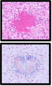
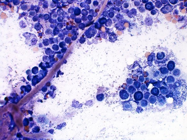
Figure 1&2: HE (top) and Gram stain (bottom). Clusters of bacteria (gram negative on gram stain) surrounded by Splendore-Hoeppli material in a lump removed from the face of a cow with suspected Actinobacillus lignieresii infection.

  • Skin biopsies were received from a five-day-old calf showing signs of flaking/scaly skin. Histopathology identified a hyperplastic and perivascular dermatitis. This is a relatively common cutaneous reaction to a wide range of insults and is not aetiologically specific. However, other changes seen included compact orthokeratotic hyperkeratosis forming mats overlying the epidermal surface and a suspicion of disorderly or abnormal hair follicle growth, which could be considered compatible with a mild form of congenital icthyosis. Typical parakeratotic hyperkeratosis as seen in zinc deficiency was not seen and therefore considered less likely in this case. Congenital icthyosis is a rare, inherited genetic disorder in cattle and can occur in severe, lethal forms or milder cases such as this.

Systemic and miscellaneous disease

  • There were several cases of suspected mucosal disease this month. On one farm a six-month-old calf was off feed, had mucosal erosions in the oral cavity and abnormal ‘gloopy’ faeces. The animal tested positive for BVD antigen with a high ‘OD’ (Optical Density) in the ELISA test confirming BVD viraemia. In another case a four-year-old Highland cow, which was showing signs suspicious of reticuloperitonitis, was found to be BVD viraemic in the antigen ELISA test and was seronegative for BVD antibody. The cow had produced a confirmed BVD PI calf the previous year but was reported to have had a previous negative result for BVD virus by tissue tag testing. Given the history and clinical signs, it was thought likely that this was a PI animal which had either a false negative result on tissue testing or had been missed out of the PI hunt in error.
  • Postmortem tissues were received from a 20-month-old bullock which had presented with neurological There was a reduced palpebral reflex, and no menace response was present. When walking the animal was unsteady, ataxic and was circling and head pressing. Diarrhoea was also observed. Grossly the liver appearance was reported as ‘very abnormal’. Histopathology identified severe, ongoing and chronic necrosuppurative hepatitis with chronic portal-bridging fibrosis and biliary hyperplasia, which would account for signs of hepatic encephalopathy. It was suggested that the biliary hyperplasia could be the result of an ongoing inflammatory process, possibly due to ascending inflammation extending up from the bile duct, or the result of abscessation in the liver. A toxicity (e.g. ragwort, chronic copper toxicity, mycotoxicosis) could also be possible. Biliary obstruction caused by liver fluke was also considered, though no fluke were seen in the tissue sections examined. The more acute necrotising hepatitis may have been part of an ongoing inflammatory process or due to secondary bacterial infection.
  • Lung tissue was submitted from a 51-day-old pre-weaned Holstein heifer The farmer reported that six calves of the same age had died over a 10-day period. Calves presented initially with signs of pneumonia but responded poorly to treatment with tulathromycin. PCR testing of lung tissue detected Mannheimia haemolytica. Changes in lung on histopathology were consistent with a septicaemia, of which M.haemolytica may have been the cause. However, salmonellosis was considered as a further possible differential and ancillary testing for this was suggested.
  • Mycoplasmopsis bovis (formerly Mycoplasma bovis) was detected in two samples of joint fluids from calves on the same farm. The vet reported that there had been multiple cases of joint effusion and lameness in calves of six to eight weeks of age. There was no evidence of navel infection but postmortem inspection of the lungs of an affected calf identified purulent material in the cranial middle and accessory lung lobes, suggestive of bacterial involvement. Changes seen in the lung tissue on histopathology included a prominent lymphoplasmacytic peri-airway infiltrate with follicle formation, areas of atelectasis, some bronchiolitis fibrosa obliterans and fibrosis of airways, frequent large multinucleated cells, and a lesser suppurative bronchopneumonia. The lymphoplasmacytic infiltrate was considered strongly suggestive of a Mycoplasmopsis spp infection, in this case most likely Mycoplasmopsis
  • Streptococcus dysgalactiae was isolated from a sample of joint fluid from a calf with signs of umbilical vein abscessation and septic arthritis
  • Mycoplasmopsis bovis was detected by PCR testing of a swab from the right ear of a six-week-old Limousin calf with mild pyrexia and head tilt of 48hrs Thick purulent material was observed in the ear canal when swabbing. Otitis rarely occurs in isolation in cases of M.bovis infection but often because of spread from the respiratory tract via the eustachian tubes to the middle and inner ear.
  • A yearling heifer showed signs of pitting brisket and ventral Mucous membranes were noted as pale on clinical examination and there was pyrexia (39.90C). Blood was taken for further investigation and haematology testing revealed a significant lymphocytosis of 20.2×10^9/l (RI = 2.5-7.5×10^9/l). A Pathologists Haematology Review (PHR) was requested to investigate the abnormal findings. Blood film examination confirmed moderate lymphocytosis with a predominance of small to medium lymphocytes. Lymphocytosis may be due to chronic inflammation or lymphoproliferative neoplasia (for example chronic lymphocytic leukaemia or leukaemic lymphoma).
  •  

Sheep and goats

Abortions, reproductive disease, mastitis

  • Multiple cases of enzootic abortion were reported again this month, primarily diagnosed on MZN-stained smears of submitted placentae, but occasionally on PCR. Abortions typically occurred in late gestation with multiple abortions occurring in some flocks. In one flock of 400 ewes, approximately 40 lambs had been aborted in the four weeks prior to lambing.
  • Multiple cases of Campylobacter fetus associated abortion were reported again this month. Where reported, most occurred in late gestation and some cases involved full term, stillborn lambs.
  • In one flock where six out of 120 ewes had aborted one to three weeks before lambing, Campylobacter jejuni was isolated from one of two samples of foetal stomach contents received.
  • Several cases of abortion associated with Toxoplasma gondii were reported, diagnosed either on PCR of placenta or foetal fluid In one flock, it was the first abortion of the lambing period and the aborting ewe had two live lambs and one dead foetus. The flock was not vaccinated for Toxoplasma.
  • Abortions associated with Listeria monocytogenes were reported, typically associated with feeding on soil contaminated or spoiled silage, or grazing on muddy ground. In one flock of ewes housed for lambing, the abortions were linked to a new source of forage.
  • In one case where Salmonella 61:k:1,5,7 was isolated from foetal stomach contents, there had been an increase in late abortions or rotten stillborn
  • A moderate growth of coli was isolated from the stomach contents of a lamb aborted at term in one flock. E. coli is typically associated with sporadic abortions, and it can also cause ‘rotten lamb syndrome’.
  • An unusual isolate from the stomach of an aborted foetus this month was Actinobacillus seminis. This is an occasional cause of abortion in sheep that more commonly causes epididymitis and orchitis in rams as an ascending infection from the prepuce, where it is an inhabitant. It is possible that infection may spread venereally.
  • Another unusual isolate was Mannheimia haemolytica, which has been reported as an occasional abortifacient in sheep.
  • Border disease viraemia was confirmed in ataxic and shaking lambs in at least three In one
  •  

Respiratory disease

  • Samples of lung received for histopathology from a two-year-old Devon Closewool ewe had an extensive and severe bacterial pneumonia consistent with the isolation of Pasteurella multocida in pure, heavy growth from a lung swab. There were changes suggestive of Mycoplasma ovipneumoniae involvement, and so this may have been a case of atypical/enzootic pneumonia. Some underlying airway damage was seen and a previous viral insult (e.g. RSV, PI3) was considered possible. A similar case was reported in three to four-week-old lambs in a second flock.
  • Post-mortem tissues were received from a crossbred, in-lamb ewe that became ill thrifty and died with copious fluid coming from the nostrils. PME revealed areas of consolidation and pallor within the lungs and OPA was confirmed on histopathology (figure 3). Infection normally occurs at a young age with progressive neoplastic transformation of pulmonary tissue and the onset of clinical signs normally from 2-3 years of age. Animals commonly present with exercise intolerance and ill thrift however if a secondary bacterial pneumonia develops this can often lead to sudden death.

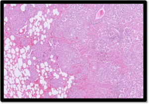
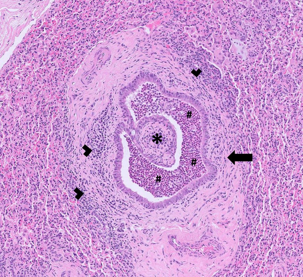
Figure 3: Neoplastic proliferations seen in the lung of a sheep with OPA

Gastrointestinal disease

  • Several cases of watery mouth were confirmed on coli culture and antibiotic sensitivity testing.
  • Several cases of lamb dysentery were suspected with the finding of Clostridium perfringens beta and epsilon toxins on the ELISA in small intestinal contents and, in one case, In the latter case, five-day-old lambs lambed indoors were turned out with the ewes and started scouring with some blood present in the faeces. The ewes had not been vaccinated with clostridial vaccine.
  • Type D clostridial enterotoxaemia was suspected on detection of Clostridium perfringens epsilon toxin in a faeces sample from a three-year-old male pygmy goat with lethargy and scour containing mucus and blood.
  • Cryptosporidiosis was diagnosed on several occasions and rotavirus type B infection on a few occasions in young lambs with scour.
  • Parasitic gastroenteritis was reported in goats and In some cases, strongyle egg counts were high enough to raise the suspicion of haemonchosis. Nematodirosis was also diagnosed in four to six-week-old scouring lambs.
  • A ram with soft faeces and bloat was diagnosed with fasciolosis on liver fluke coproantigen ELISA. The ram had received a combination drench containing triclabendazole 18 days earlier, consistent with treatment failure. Fasciolosis was also diagnosed on coproantigen ELISA in a group of eight pet goats on permanent grazing which were all in poor condition; the faeces sample also had a strongyle egg count of 1050 epg, consistent with concurrent parasitic gastroenteritis.
  • Several cases of Johne’s disease were diagnosed in sheep and goats through serology and faecal PCR, typically presenting with weight

Skin and eye disease

  • Seroconversion to sheep scab was seen in sheep with clinical signs including exudative dermatitis, wool loss and pruritus.
  • Haired skin samples were received from the third lamb born dead in a flock with a skin condition where the wool was compacted into soft clumps and peeled off the skin easily. Histopathology detected severe, diffuse, hyperkeratosis with hair follicle dysplasia, suggesting a diagnosis of ichthyosis fetalis, a non-epidermolytic Ichthyosis is a heterogeneous group of rare congenital, often autosomal recessive, disorders affecting skin cornification and keratinisation, characterised by an excessive accumulation of superficial scales on the skin.

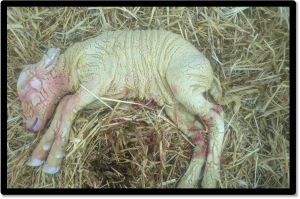
Figure 4: Ichthyosis fetalis in a lamb. Photo courtesy of Emily Haydon and Violeta Lazaro Alvarez, Drove farm vets.

  • Mycoplasma conjunctivae infection was diagnosed on PCR testing of plain ocular swabs in at least two flocks, and, in both cases, there was difficulty getting on top of M. conjunctivae infections regularly recur due to the difficulty in eradicating the bacterium from deep within the conjunctival sac.

Systemic and miscellaneous disease

  • Several cases of CLA were diagnosed primarily in goats on culture and serology. In one goat herd, a buck and a few does were bought in from a supposedly CLA tested negative The buck and one doe developed multiple lumps in the region of mandibular and prescapular lymph nodes, with an abscess on the buck bursting. A swab from the affected doe was positive on culture and one of four other goats blood sampled was seropositive for CLA.
  • Staphylococcus aureus was isolated from a cheek abscess on a three-year-old Boer
  • A large sheep enterprise had 30 lambs die between five days and three weeks of age, with some found dead but the majority found recumbent, bloated and some with scour, and all dying within one One lamb was examined postmortem; the lamb was in good body condition and the only finding of note was mild pericarditis. Epsilon toxin was detected on Clostridium perfringens toxin ELISA, most likely consistent with pulpy kidney although brain histopathology is required to confirm the diagnosis. The ewes were not vaccinated against clostridial disease. Cases were also reported in lambs under two weeks old dying acutely in two further flocks.
  • Streptococcus dysgalactiae was isolated from a lamb of less than a week of age in one flock and in a less than three-week-old lamb in a second This is the most common cause of joint ill in young lambs.
  • A carpal joint fluid aspirate was received from a seven-day-old Welsh Mountain lamb with joint ill that had failed to respond to A scant growth of E. coli was isolated which was resistant to amoxycillin but sensitive to potentiated amoxycillin.
  • Samples were received from one of several lambs to have died in the first week of life in an 800-ewe flock. On histopathology, there was evidence of necrosuppurative pneumonia and hepatic abscessation, and lesion character and distribution were consistent with embolic spread due to sepsis. Listeria, Campylobacter and Fusobacterium necrophorum were all suggested as potential causes.
  • Post-mortem tissues were received from a sheep, after several periparturient ewes died following the development of neurological signs that were unresponsive to magnesium, calcium, penicillin, vitamin B1 and NSAIDs. On histopathology of brain, there was severe, suppurative, granulomatous and plasmalymphocytic, necrotising, rhomboencephalitis and meningitis which was highly suggestive of listerial encephalitis due to Listeria monocytogenes. A second case was diagnosed with brain histopathology in a four-year-old Texel cross ewe in a flock in which several sheep had developed
  • Orf was confirmed as the cause of ‘sores and wounds’ on the mouth and tongue of Romney sheep – nasopharyngeal swabs taken from four sheep were all positive on PCR.

Camelids

  • Eimeria macusaniensis was detected in a faeces sample from a very thin alpaca cria with a tucked-up E. macusaniensis may cause a severe necrotising enteritis in alpacas.
  • Parasitic gastroenteritis was diagnosed in an adult female alpaca with condition loss that had a strongyle egg count of 1050 epg. 200 epg Trichuris sp. and 50 epg Capillaria sp. were also detected, and these gastrointestinal nematodes are also known to be pathogenic in South American camelids.
  • Fasciolosis was confirmed in a female alpaca on liver fluke coproantigen

Skin and eye disease


  • Culture of a swab taken from multiple infected ulcerative lesions on the eyelid of a seven-year-old male alpaca grew a profuse, pure growth of Mammaliicoccus sciuri. This is a known opportunist pathogen in humans and pigs, and the lesions were visibly irritating, the alpaca rubbing its eyes on the fence.
  • A two-year-old alpaca that was otherwise in good condition and was systemically well, developed ulcerated, raised lesions over the hind legs (circumscribed) and chest (diffuse), raising concerns about a neoplastic The histological appearance was of very large numbers of mast cells within the dermis, accompanied by eosinophils, and with superficial crusting with secondary bacterial infection. It was unclear whether this represented a reactive mast cell population with eosinophils, or a neoplastic process. Mast cell neoplasia in alpacas is extremely rare, although mast cell tumours have been reported in a llama, appearing as multiple cutaneous masses. A hypersensitivity response was therefore considered to be a potential underlying cause, although the stimulus was not identified.

Systemic and miscellaneous disease

  •  Blood samples were received from a six-year-old alpaca that was losing weight and anaemic, having a PCV of 14% two weeks earlier which had not responded to an iron injection, the PCV being 8% just before the samples were A PCV of 16% was obtained at Axiom, the anaemia was regenerative (reticulocytes 107 x 109/l) and Mycoplasma haemolamae was detected by PCR. M. haemolamae infection typically manifests as a regenerative anaemia, often in animals that are stressed, immunosuppressed or have concurrent illnesses.
  • Tissues were received from a five-year-old male neutered alpaca that had a large mass on the outer toe which was removed surgically. Histopathology revealed spindle cell proliferation and a benign fibroma was considered the most likely diagnosis. However, it was not possible to completely rule out a low-grade sarcoma.
  • Schaalia (formerly Actinomyces) was isolated from a cheek abscess in an alpaca. Schaalia sp. are opportunistic pathogens associated with infections such as abscesses and actinomycosis, and so it was likely to be a significant finding in this case.

Other Species

  • Five of a group 10 seven-week-old pigs in a litter died after over a two-week history of hind limb ataxia and anorexia progressing to clonic seizures. The remaining pigs were also looking thin but the litter in the neighbouring pen were unaffected. A heavy pure growth of Streptococcus suis was isolated from a swab of brain suggesting this was a case of suis associated meningitis.
  • A four-year-old male reindeer died after developing pyrexia and respiratory signs, despite supportive treatment. On PME, there was blood-tinged pleural fluid, bilateral marked pulmonary congestion and evidence of rumenitis. Histopathology was consistent with a fibrinosuppurative bronchointerstitial pneumonia consistent with a bacterial bronchopneumonia with septicaemia

Deficiencies and toxicities

  • A Jersey cow which had been calved for a few weeks and was reported by the owner as ‘failing’ was found on blood testing to have a high BHB level of 2mmol/l (0.00-1.2mmol/l) indicative of ketosis, a high NEFA level pointing to excessive fat mobilisation and a severely raised GLDH of 1192 U/l (RI 0-25U/L) consistent with severe hepatocellular damage. The hepatopathy was suspected to be due to fatty liver disease, though other causes of hepatic injury could not be excluded. In this case the diet was found to be inadequate for the cows’ energy and protein needs and improvements had been initiated.
  • Evidence of copper and selenium deficiency was identified on trace element screening of a group of Aberdeen angus cross cows which were failing to show signs of oestrus. They had been on poor grazing with calves at foot and were receiving no supplementation. All seven cows tested had very low copper levels ranging from 2.4-5.7µmol/l (RI 9-19µmol/l) and low GSH-Px levels ranging from 12-29 U/L RBC (RI>30 U/L RBC).
  • Evidence of selenium deficiency was identified in a suckler herd on a forage only diet which were producing small, weak calves. All six animals tested had low GSH-Px levels consistent with selenium deficiency. All the tested cows also had low or suboptimal urea levels indicating a lack of rumen degradable protein most likely due to low levels of RDP in the diet in this
  • A group of Hereford cross cattle grazing coastal land were reported to be performing poorly with cows in poor body condition, not conceiving and with some skin sores over their Some of the store cattle were failing to gain weight as expected. Ragwort had been present and was picked but not cleared from the fields. Trace element testing identified hyposelenosis in five of the six tested animals. Urea levels were very low in all cows (below the lower detection limit of the test of 0.8mmol/l) indicating there was insufficient rumen degradable protein in the diet. In several of the animals albumin levels were below optimum suggesting the possibility of long-term low protein supply, underlying parasitism or hepatic disease. Three cows did have mild to moderately raised GLDH levels indicative of some hepatocellular damage of which ragwort ingestion is one possible cause. All six tested cows had raised pepsinogen levels with four of these being above the 2.5iu/l level indicating a level of abomasal damage due to parasitism likely to be associated with clinical disease.
  • There was evidence of vitamin A deficiency (Vitamin A = 218ug/l RI = 249-500ug/l) in a six-week-old calf which was showing signs of poor growth, lethargy, alopecia of the ears and absence of a menace
  • Vitamin E deficiency was suspected to be the cause of a stiff gait, muscle weakness ataxia in a three-week-old lamb with a low vitamin E level of 0.9mg/l (RI = >1.0mg/l).

Follow us on Facebook

https://www.facebook.com/Axiomfarmvetlab/

This newsletter is sent by e-mail to each vet practice but if you would like a copy sent to your individual e-mail account please contact us at dsfarm@axiomvetlab.co.uk and we can add you to our circulation list.

Related Posts