Farm May Newsletter 2026

Dedicated farm line – 01626 357776

Follow us on Facebook

https://www.facebook.com/Axiomfarmvetlab/

This newsletter is sent by e-mail to each vet practice but if you would like a copy sent to your individual e-mail account please contact us at dsfarm@axiomvetlab.co.uk and we can add you to our circulation list.

Would you like to send the Axiom farm vets photos of PMEs to allow us to assist with sample selection and diagnosis? Please send photos by WhatsApp to 07944 649222.

Email us with your additional test requests

You may find this is a more efficient way of making requests than phoning the farm team, saving you time in your busy day. Our farm team also find it a more efficient way of dealing with your requests.
The email address for test requests is: dsfarm@axiomvetlab.co.uk 

Axiom Farm Vets

If you wish to speak to a particular farm vet about one of your cases, you can consult the table below to see when our vets are usually available. A duty farm veterinary advisor is always around on a Saturday morning from 9am-12pm.

Farm Vets

Axiom Farm Factsheets

Did you know there are lots of farm animal factsheets available on the Axiom Veterinary Laboratories website? These can be downloaded and used as handy guides when making decisions on sampling and testing. The factsheets can be accessed using the link below or scan the QR code in the following article on farm histopathology: Farm Animal Fact Sheets Axiom Veterinary Laboratories

Farm histopathology

We offer farm histopathology on postmortem samples under the code FPM01. Up to three different tissues can be processed under this code and extra tissues can be processed on request. Where different lesions/ stages of disease are seen in one organ (e.g. pneumonic lungs) multiple small sections can be submitted as one tissue type and this will not incur additional costs. For best results only perform histopathology on tissues from fresh carcases and ensure samples are transferred into fixative as soon as possible after sampling. Scan the QR code below to access our ‘How to get the most out of your large animal histopathology service’ factsheet. Further tips on sampling for histopathology can be found in the ‘Histopathology: points to consider’ and ‘Sampling for respiratory disease’ factsheets.

Feedback on our services

As part of our continuing effort to help improve our services to clients within the Laboratory Division we have put together a short survey which will be sitting live permanently for anyone to complete whenever they have a few minutes spare. This can be filled in by anyone who uses any of our services and we encourage feedback to help us understand what we are doing well and where we need to make improvements. The QR code and link to the survey will remain live as a continuous tool to enable us to always gather feedback. If anyone has any questions about this, they are welcome to contact either of the Quality Managers at the Laboratories, Claire Richardson for Axiom Veterinary Laboratories and Susan Reeve for Finn Pathologists. Thank you in advance for helping us to improve our services.

https://www.surveymonkey.com/r/Laboratory_Satisfaction_Survey

BTV testing guidance

We wish to remind vets that as a commercial laboratory we are unable to offer any Bluetongue testing either on blood samples or abortion material. If we receive samples from cases in which Bluetongue is suspected, we will be required to safely dispose of the samples without further testing. We are always happy to discuss cases over the phone before submission of samples. Clinical suspicion of BTV infection must be reported to APHA.  Further information on investigating poor reproductive performance in cattle and sheep during Bluetongue outbreaks can be found at the following link: Bluetongue outbreaks (accessible version) – GOV.UK

Molecular mastitis test

Unfortunately, the Mastitis PCR referral test is no longer available to us. As an alternative you may wish to consider the Mastitis Rapid Molecular test which is available at Axiom (Test code: FAMAS). This test detects the presence of six key mastitis pathogens, and the turnaround time is no longer than the following working day. This can allow a targeted course of treatment based upon the pathogens present, aiding responsible use of antibiotics and resulting in less discarded milk. As well as potentially saving money there are benefits for herd health in the long term. The agents detected are E. coli, Staphylococcus aureus, Streptococcus uberis, Streptococcus dysgalactiae, Streptococcus agalactiae and Klebsiella pneumoniae. The cost of the test is £45 & VAT (test code FAMAS).

Interesting cases in March

Cattle
Abortions/mastitis

  • Listeria innocua was isolated from a foetal stomach content in one case of bovine abortion from a herd which had a recent history of premature and stillborn calves. Although generally considered as non-pathogenic, some Listeria innocua isolates have recently been shown to possess virulence factors which could result in significant pathogenicity and potential for disease. Water troughs and soil can be potential
  • There were several abortions associated with Listeria monocyotogenes this month, all of which occurred in late gestation.
  • A moderate growth of Aspergillus terreus was isolated in fungal cultures of foetal stomach contents from a bovine foetus which had been aborted in the seventh month of A.terreus is recognised as a cause of bovine mycotic abortion and the finding was likely to be of significance in this case.
  • A heavy and pure growth of Trueperella pyogenes was cultured from a bovine foetus aborted in mid-gestation. pyogenes is a known cause of abortions in cattle. In this case the submitting vet noted that there were no visible gross lesions on examination of the foetus, which is very common in abortion investigations.

Respiratory disease

  • Severe lung pathology was observed during postmortem examination of two Aberdeen Angus cross calves which were from a group of thirty affected by respiratory disease. RSV and PI3 virus were detected by PCR in lung tissue in one of the RSV, but not PI3, was detected in the sample from the other animal. RSV was also detected in a pooled test of nasopharyngeal swabs from acutely affected live calves. Mycoplasmopsis bovis (formerly Mycoplasma bovis), Histophilus somni and Pasteurella multocida were also detected in lung tissue from both calves. In addition, Mannheimia haemolytica was detected by PCR in the nasopharyngeal swabs but was not detected in lung tissue.
  • A five-year-old dairy cow presented as a sudden death case, six days after infusion of a dry cow intramammary At postmortem examination there was evidence of mastitis in one back quarter. However, the main postmortem finding grossly was extensive, severe cranioventral consolidation and caudodorsal emphysema of both lungs. The trachea was described as severely inflamed. Cultures of lung tissue produced a mixed growth of contaminants. PCR testing of tissue detected IBR virus, Mycoplasmopsis bovis, Histophilus somni and Pasteurella multocida. The appearance on histopathology was of a chronic suppurative pneumonia with abundant bacteria. In the lung sections examined there was no convincing evidence to suggest Mycoplasmopsis bovis or pneumotropic viral involvement.
  • Post-mortem tissues were received from a five-month-old, post-weaned, Angus cross, beef finishing heifer Ten calves of a group of 100 had died following respiratory illness. The vaccination history of the group was unknown. The lung lesions observed at postmortem examination were recorded as suspicious of Mycoplasmopsis bovis infection by the submitting vet. Bacteriology of a transport swab of lung was unrewarding. PCR testing for respiratory pathogens detected Mycoplasmopsis bovis, Histophilus somni and Pasteurella multocida. Histopathology identified extensive, severe, chronic-active, plasmalymphocytic and suppurative bronchointerstitial pneumonia with bronchiolitis fibrosa obliterans. The changes were considered suggestive of enzootic calf pneumonia/bovine respiratory disease (BRD). However, due to the chronic and established nature of the lesion, it was not possible to confirm or rule out any underlying pathogen. Bronchiolitis fibrosa obliterans indicates a severe necrotising bronchiolitis and in this age of calf is normally associated with a previous pneumotropic viral infection such as BHV 1, RSV and PI 3 (though virology may have been unrewarding at this stage due to the chronic nature of the lesions). There was evidence of secondary bacterial infection consistent with the detection of Pasteurella multocida, and Histophilis somni. In addition, a plasmalymphocytic peri-airway infiltrate indicated likely concurrent mycoplasma infection consistent with the Mycoplasmopsis bovis.
  • Pasteurella multocida was isolated in cultures and detected by PCR in lung tissue from an eight-week-old calf which had shown signs of pneumonia from three days of age. Chronic bronchopneumonia with bronchiolitis fibrosa obliterans and fibrous pleurisy was observed on histopathology. The changes were likely to reflect previous damage to the lung which may be mediated by viral, mycoplasmal and bacterial causes or a combination. At this late stage in the disease the underlying cause of the damage was In this case the P.multocida isolate demonstrated invitro resistance to multiple antibiotics, which may have reflected various treatments during the life of the calf.
  • Postmortem tissues were received from a four-year-old cow which was the second in the herd to recently show signs of milk drop and weight loss in early pregnancy. The lungs were reported to have appeared congested with emphysematous bullae and abscesses throughout, and a purulent discharge was present in the trachea. Trueperella pyogenes was isolated in cultures of lung tissue. Histopathology identified a multifocal, moderate, subacute suppurative bronchopneumonia with intralesional bacteria and intravascular thromboembolism. This morphological diagnosis along with the gross description of the lungs and isolation of pyogenes suggested the most likely cause was embolic pneumonia. This is often the result of spread from an inflammatory focus elsewhere which could include e.g. liver abscess, endocarditis, metritis, mastitis or foot abscess.
  • Inhalation/aspiration pneumonia was suspected to be the cause of death in a calf aged between one and two Histopathology identified a multifocal, subacute, mild-moderate, necrosuppurative bronchopneumonia with frequent multinucleated giant cells with intracytoplasmic inclusions and diffuse atelectasis. These findings were considered unusual. Frequent multinucleated giant cells containing eosinophilic intracytoplasmic inclusions were seen in the alveoli. Similar intracytoplasmic eosinophilic inclusions were occasionally seen in airway epithelial cells. These are features most often seen with RSV or PI3 infections, though other changes associated with these pathogens were not present in this case. Viral PCR was suggested to further investigate possible viral involvement. There was also evidence of suppurative bronchopneumonia and necrosis indicating secondary bacterial infection. It was suggested that the multinucleate giant cells may have been engulfing foreign material or fibrin resulting from inhalation / aspiration possibly following tube feeding.

Skin and eye disease

  • Listeria monocytogenes was isolated in cultures of eye swabs from a cattle herd in which multiple animals were affected with bilateral ophthalmitis. The finding was considered significant and consistent with a case of bovine iritis (silage eye)
  • Moraxella bovoculi was isolated from an eye swab in a case of Infectious Bovine Keratoconjunctivitis (IBK). The finding of Moraxella bovoculi is considered significant as a cause of eye disease in cattle and is often considered as the primary cause of this However, it can also be found in healthy eyes and likely works synergistically with other factors in cases of eye disease.
  • A cutaneous mass from an eighteen-month-old British Blue cross heifer was identified as a melanocytic tumour (melanoma) by histopathology. Melanomas in cattle are often benign and the neoplastic cells in this case appeared to have a low mitotic count which would be supportive of a benign tumour. However, due to only a small part of the mass being submitted for examination it was not possible to assess for local infiltration into surrounding tissues and malignancy could not be ruled out based on the sections received. It was advised that if the mass clinically appeared well demarcated or was on a stalk, then that could potentially further support it being a benign tumour.

Systemic and miscellaneous disease

  • BVD viraemia was detected by PCR testing of serum from a seven-day-old female calf described as a ‘dummy calf’, with a dome headed appearance and showing signs of ataxia. BVD virus can cause severe damage to the foetal brain leading to hydrocephalus or cerebellar hypoplasia resulting in an abnormally shaped skull and incoordination of movements respectively.
  • Mucosal disease was suspected in a six-month-old heifer which tested strongly positive for BVD virus in the antigen ELISA It was the second animal in a group to show signs of a watery scour, mucopurulent nasal discharge and inappetence. The first affected animal had died within a few days of first showing signs. Outbreaks of mucosal disease can occasionally occur if there are multiple PI animals in a group.
  • A cow with suspected renal issues died and when examined postmortem there was gross pathology of one of the kidneys with a dilated renal pelvis and crystalline material visible within. A marked, subacute and necrotising pyelonephritis characterised by necrosis and cavitation, with some evidence of mineralisation and large numbers of associated bacteria was identified by histopathology. The appearance was suggestive of ascending infection from the lower urinary tract. Typically, in these cases, only one kidney is affected. The mineralisation was likely to be a result of necrosis and inflammation of the kidney tissues. Cystitis and pyelonephritis are seen in cattle, often soon after calving. Corynebacterium renale is the pathogen most associated with the condition, although bacterial cultures were not performed in this case.
  • Postmortem tissues were received from a six-month-old Aberdeen Angus cross heifer, which was reported to have become unthrifty over a short period of At postmortem examination the carcase appeared mildly jaundiced and the liver was abnormal. The heart appeared enlarged. Histopathology of liver identified sheets of neoplastic cells with scant cytoplasm and round-ovoid nuclei with dense chromatin, surrounding and expanding portal tracts and forming coalescing clusters. In the heart clusters of round neoplastic cells infiltrated and separated cardiac myofibres. Changes were considered consistent with round cell neoplasia, most likely a lymphoma. Sporadic juvenile lymphoma is seen in calves usually around 3-6 months of age and can be present from birth. Cases are rare and typically present with poor body condition and enlargement of lymph nodes.

Sheep and goats

Abortions, reproductive disease, mastitis

  • Numerous cases of enzootic abortion were diagnosed in sheep flocks, primarily on MZN-stained smears of placentae but also on Chlamydia abortus Where a history was given, many affected flocks had experienced multiple abortions in the last few weeks of pregnancy, some flocks also had still born, weak and poor doing lambs and there was evidence of placentitis.
  • Numerous cases of Campylobacter fetus associated abortion were reported in sheep flocks, with multiple abortions being reported in the last four weeks of gestation. Occasional still births and weakly lambs were also reported and ewes were typically well in themselves.
  • A few cases of Campylobacter jejuni associated abortion were also reported, with multiple abortions.
  • Numerous cases of Toxoplasma gondii associated abortion were diagnosed on PCR or foetal fluid Multiple abortions were reported in many affected flocks and in some cases weakly lambs and mummified lambs were seen.
  • Numerous cases of abortion associated with Salmonella 61:k: 1, 5,7 were diagnosed in sheep flocks. This is a variant of the sheep adapted strain of Salmonella enterica diarizonae. In some cases, aborting ewes were reported to be sick and/or scouring.
  • Salmonella Montevideo was isolated from the stomach contents of aborted foetuses in two flocks. This isolate has been associated with gulls.
  • Several cases of abortion associated with Listeria monocytogenes were reported. This is typically associated with the ingestion of spoiled or soil contaminated silage or grazing on muddy ground.
  • In several flocks, coli was isolated in pure growth from the stomachs of aborted foetuses. Typically, E. coli causes sporadic abortions associated with opportunistic invasion of the gravid uterus but, in some flocks, multiple abortions were reported.
  • Other potential causes of opportunistic infection were reported where a less common potential abortifacient was isolated in pure growth from foetal stomach contents. These included Streptococcus dysgalactiae, Streptococcus uberis, Acinetobacter pseudolwoffi and an Aeromonas species.
  • Border disease viraemia was confirmed in weak, shaking and dead lambs in at least three flocks. Exposure to BDV was also confirmed on serology in several flocks with poor fertility and high barren
  • Mastitis cases this month included Staphylococcus aureus in three goats from different holdings, and Mannheimia haemolytica and coli, each from two ewes. An unusual isolate from the milk of a Dutch spotted ewe was Corynebacterium pseudotuberculosis, the causative agent of CLA. This has been reported, for example, when an abscess forms within the mammary gland.
  •  

Respiratory disease

  • Several cases of pneumonia were diagnosed in lambs, including cases of bacterial bronchopneumonia where Mannheimia haemolytica was suspected to be implicated and, in one case, successfully Cases of a more chronic nature which were suspected to be multifactorial in origin with a possible underlying viral and/or mycoplasma involvement, and cases of bronchointerstitial pneumonia which were considered to be part of a septicaemia.
  • A nasopharyngeal swab was received from a four-year-old Lincoln Longwool ram with a history of chronic pneumonia over three to four Clinical examination found constant bilateral copious white-green nasal discharge. Salmonella enterica ssp. diarizonae serovar 61:k: 1, 5,7 was isolated, a known cause of chronic rhinitis in sheep which usually fails to respond to antibiotic treatment.

Gastrointestinal disease

  • Rotavirus Type B was detected by PAGE in faeces from scouring lambs in two flocks. In one flock, 60-70% of lambs were scouring despite good colostrum management and good hygiene.
  • Type D clostridial enterotoxaemia was suspected in an adult goat with scour after the detection of epsilon toxin on Clostridium perfringens toxin ELISA testing of faeces. This was the only adult goat affected but most of a group of 140 six-month-old goats on the farm had scour and 25 had It was also diagnosed in a five-year-old goat that was collapsed in a field, inappetant and, on clinical examination, had white mucous membranes, tachycardia and slow rumen turnover.
  • Numerous cases of Johne’s disease were diagnosed in sheep and goats on serology and faecal Where clinical history was provided this include weight loss, bottle jaw, scour and inappetence.
  • Salmonella enterica diarizonae serovar 61:k: 1, 5,7 was isolated from the faeces of a six-week-old lamb with scour. This sheep adapted strain has been associated with enteritis in sheep.
  • Several cases of fasciolosis were diagnosed on fluke microscopy and liver fluke coproantigen ELISA testing in sheep this month, including a thin ewe, one month off lambing, that was bright but thin, recumbent and scouring.
  • Several cases of parasitic gastroenteritis were diagnosed on faecal egg counts in sheep and Some animals with counts in the thousands were suspected of having haemonchosis, for example a pooled sample from three anaemic ewe lambs with a BCS of 1/5 had a strongyle egg count of 8,000 epg.

Skin and eye disease

  • Animals in several flocks were seropositive for sheep scab. Where clinical signs were reported, these included wool loss, skin scabs and pruritus. In some cases, no Psoroptes mites had been seen in skin.
  • A mixed Dermatophilus congolensis and Orf infection was diagnosed on PCR testing of samples of lesions from one of two one-year-old ram lambs with proliferative lesions on the head and legs (figure 1). Orf is likely to be the primary infection in these cases, known colloquially as ‘strawberry footrot’.  Orf was also confirmed on PCR testing in a one-year-old Texel ram with extensive scabs and pustular lesions over the head.

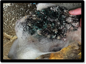
Figure 1: Proliferative lesions on the head of a ram lamb with mixed Orf and Dermatophilus congolensis infection (Photo: Nikki Prosser, Towcester farm Vets)

  • Mycoplasma conjunctivae was confirmed on PCR testing of pooled plain ocular swabs as the cause of keratoconjunctivitis in a sheep flock in which around 20 animals were affected.

Systemic and miscellaneous disease

  • Seroconversion to Maedi visna was reported in several flocks. Cases included a winter shorn mule flock with ongoing 3-4% ewe mortality following chronic respiratory signs and loss of body condition, in which five out of six animals sampled were seropositive, and a re-accreditation screen of recipient lambs entering an MV accreditation flock, where seven out of 167 were seropositive.
  • After two cases of CLA were diagnosed in a goat herd (one on culture, one on serology), a further 11 animals were screened using serology and 9 animals were One of the seronegative goats had a visible parotid abscess from which C. pseudotuberculosis was isolated; abscesses can be detectable from eight to nine days following infection; however, seroconversion can take two weeks. In CLA-affected herds, clinical signs and bacteriology should therefore take precedence over serology in the diagnosis of infection.
  • Pulpy kidney was suspected when epsilon toxin was detected by ELISA in the intestinal contents of one of two two-week-old lambs that died after a few hours of bloating. The lambs were not vaccinated for clostridial disease. Brain histopathology is required to confirm diagnosis.
  • Streptococcus dysgalactiae was isolated from the joints of several lambs in different This is the most common cause of joint infection in young lambs. In one 60-ewe flock, housed for three weeks and that had been lambing for one week, six lambs had become limp and weak over a two-day period. On PME, one lamb had polyarthritis and the second had fibrinous pericarditis and pleurisy. S. dysgalactiae was isolated from swabs taken from a joint of the first lamb and the pericardium of the second lamb, suggesting the latter had S. dysgalactiae associated bacteraemia. Both lambs also had low vitamin E levels. The main source of vitamin E for neonatal lambs is colostrum so low results can be due to insufficient colostral absorption. Alternatively, the dams could be deficient, but testing of ewes would be necessary to determine if the latter was the case. Colostrum quality was reported to be adequate in the flock.
  • Histopathology confirmed a diagnosis of cerebral listeriosis in a sheep with the finding of severe asymmetrical rhombencephalitis (Figure 2).

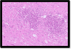
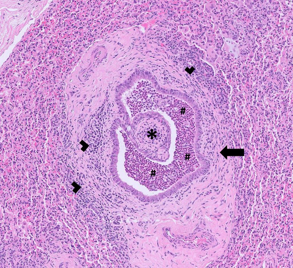
Figure 2: Microabscesses (clusters of neutrophils) seen in the brain of a sheep with listeriosis.

  • Granular cell lymphoma was diagnosed on cytology of a fine needle aspirate taken from a mass in the prescapular region of a male neutered pet sheep (figure 3). Lymphoma is not uncommon in sheep, typically arising as sporadic cases in older animals. Granular lymphoma is well-recognised in dogs and cats but is not widely reported in ruminants. In other mammals, it typically arises from the GI tract, liver or spleen, and varies from indolent to aggressive behaviour, depending on the species and presentation; it is often associated with leukaemia. Granular lymphoid cells are typically T-cell in origin but can be of the null-cell phenotype. It is possible that granular lymphoma is the same entity as ‘globule leukocyte tumour’ reported in ruminants. As in cattle, lymphoma in sheep can also arise following exposure to bovine leukaemia virus (BLV), but this is considered very unlikely here.

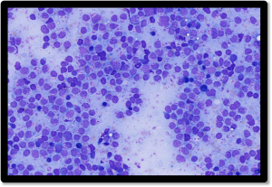
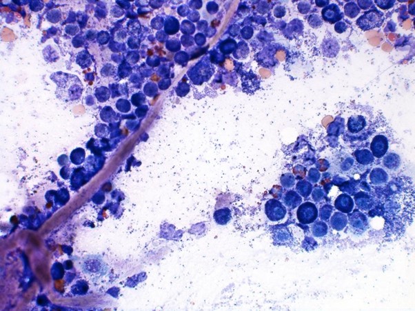
Figure 3: FNA from a mass in the left prescapular region of a male neutered pet sheep, consistent with granular cell lymphoma. Wright-Giemsa stain, x 500 magnification. There is a dominant population of atypical intermediate lymphoid cells containing numerous coarse magenta cytoplasmic granules. Occasional well differentiated small lymphocytes, plasma cells and erythrocytes are also present

Camelids

  • Fasciolosis was diagnosed on liver fluke coproantigen ELISA testing in one of two sampled camelids in a zoological collection. No clinical history was provided.
  • Parasitic gastroenteritis was diagnosed in a thin, recumbent nine-year-old male alpaca with a strongyle egg count of 750 A high strongyle egg count of 1350 epg was also found in a faeces sample from a Bactrian camel in a zoological collection; no clinical history was provided.
  • Johne’s disease was diagnosed on faecal PCR in alpacas in two different No clinical history was provided.
  • Histopathology of a slow-growing, firm lump on the mucocutaneous junction of the lip of an alpaca was consistent with a fibropapilloma.
  • Mycoplasma haemolamae infection was confirmed on PCR testing of an EDTA blood sample from a one-year-old male alpaca. No history was provided but presumably the animal was anaemic.
  • Biopsies were received from a 20-year-old female alpaca with a haemorrhagic infected mass on the right stifle. Histopathology was consistent with an epithelial neoplasm and the presence of extensive ulceration and local infiltration raised concern for malignant neoplasia. Possible differentials included basosquamous carcinoma or potentially malignant pilomatrixoma.
  • A two-year-old female alpaca developed several abscessed lymph nodes over a two-month period. Cytology on a fine needle aspirate taken from an affected inguinal lymph node detected marked neutrophilic inflammation and Staphylococcus aureus was isolated on culture. Serology for CLA was negative.

Other Species

  • A mild outbreak of scour occurred in pigs in a 70-sow farrow to finish indoor housed herd a few days after weaning, peaking at around nine to ten weeks of Three pigs were also found dead with no premonitory clinical signs, but these cases may have been unrelated. Three samples of loose faeces were submitted from recent weaners, pigs at three weeks post weaning and finisher pigs which had experienced severe scour previously. K88 E. coli was isolated from the recently weaned pigs and Lawsonia intracellularis was detected by PCR in the pigs three weeks post weaning, suggesting a mixed aetiology in the outbreak of scour.
  • Fasciolosis was diagnosed in a reindeer on liver fluke coproantigen ELISA testing following treatment for liver fluke, consistent with treatment failure.
  • A strongyle egg count of 1450 epg from a reindeer was suspected to be consistent with parasitic gastroenteritis although no clinical history was provided.

Deficiencies and toxicities

  • Pregnant ewes in good body condition which were being fed with good quality hay, sugar beet and concentrates appeared lethargic and All three ewes had high BHB levels of 5.34, 2.75 and 6.32 mmol/l (RI = 0.00-1.20mmol/l) and very high NEFA levels of 2461, 2220 and 1618umol/l (RI = 0-400umol/l) confirming ketosis and excessive body fat mobilisation. These results were indicative of pregnancy toxaemia. Although commonly associated with underfed sheep in low body condition, ewes in body condition >4 can also be at risk of pregnancy toxaemia. Reduced rumen capacity in late gestation leads to lower feed intake which in turn results in mobilisation of body fat for energy needs and fatty infiltration of the liver. Lack of exercise also appears to be a risk factor for development of the condition in over conditioned ewes.
  • Two ewes from a flock of twenty-five Soay died suddenly and the following day a further two ewes were found recumbent and morbidly Both ewes were found to have low blood calcium levels of 1.01 and 1.11mmol/l (RI= 2.00-3.00), consistent with a diagnosis of hypocalcaemia. BHB levels were within the expected reference intervals.
  • A cow which went down and started seizuring in the milking parlour and died shortly after was found to have a low level of magnesium in ocular fluid of 0.28mmol/l. Aqueous humour magnesium levels of < 0.33 mmol/l are indicative of severe magnesium deficiency, so in this case the finding was strongly suggestive of death due to hypomagnesaemic tetany (staggers).
  • Hypomagnesaemia was thought to be the cause of recumbency in a five-month-old suckler heifer, which otherwise appeared bright and alert with no other neurological signs. Serum magnesium levels were found to be very low at 0.21mmol/l (RI = 0.7-1.3mmol/l). Fast growing, milk-fed suckler calves are at risk of hypomagnesaemic tetany, which most frequently presents as hyperexcitability, seizures or sudden death.
  • Very low urea levels of <0.80mmol/l (RI= 2.0-6.6mmol/l) were seen in all five suckler cows tested from a farm which had previously reported low urea levels on a mixed diet of straw and silage and were being rechecked following a change to silage alone. It wasn’t clear from the clinical history how recently the change had taken place, though blood urea levels respond very quickly to increases in crude protein levels (within a few days). Silage, particularly that which has been late cut, matured or is low in dry matter may contain insufficient protein to meet the demands of pregnant suckler cows.
  • A one-week-old calf which was born with abnormal eyes, suspected to be congenital cataracts was found to have hypovitaminosis A. The vitamin A level in serum was 35ug/l, well below the reference interval of 249-500ug/l. Congenital eye defects including cataracts are associated with vitamin A deficiency in the dam during pregnancy, so the level in this case may have reflect low vitamin A status in the cow during ocular development of the foetus. However, as colostrum is an important source of this vitamin, if colostral intake has been inadequate, this may also be reflected in the serum levels in the neonatal calf.
  • Zinc deficiency was suspected to be involved in the development of skin lesions (crusting, scabs around the muzzle) of adult All five tested goats had serum zinc levels below the reference value of >9.9umol/l. A PCR test of skin scabs for parapox virus (Orf) was negative. Low zinc status is associated with skin disease in goats and often presents as alopecia or crusty, scaling lesions on the legs, face , ears and tail base.
  • Skin biopsies were received from a two-year-old female alpaca showing signs alopecia and skin crusting around the eyes and nose and on the limbs. Histopathology identified changes (hyperkeratosis) consistent with a keratinising disorder (figure 4) Zinc responsive dermatosis in alpacas may be due to a true zinc deficiency or a keratinising disorder that is responsive to zinc Therefore, this has also been termed idiopathic hyperkeratosis. Lesions in affected animals consist of thickened skin with tightly adherent crust that are found more commonly on hairless areas of the body such as the ventral abdomen, axilla, medial thigh and inguinal area. They can also occur on the face, pinnae and neck. Coloured animals appear to be more susceptible and young animals, typically breeding females, are more frequently affected. Dark fleeced animals tend to be more affected because darker fleeces contain high levels of zinc compared to white fleeces and exhibit higher demands on mineral metabolism. It is also important to note that zinc responsive dermatosis mainly affects one individual in a herd even when all animals are on the same diet. In this animal the zinc level in serum was at the lower end of the reference interval at 3.1umol/l (RI= 3.0-14.6). However, it is known that zinc serum levels are not considered a reliable diagnostic tool for diagnosing this condition in alpacas, as affected animals can have normal zinc levels and animals with levels below the reference interval can appear healthy.

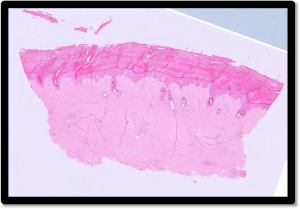
Figure 4: Marked hyperkeratosis suggestive of a zinc responsive dermatosis in an alpaca

Follow us on Facebook

https://www.facebook.com/Axiomfarmvetlab/

This newsletter is sent by e-mail to each vet practice but if you would like a copy sent to your individual e-mail account please contact us at dsfarm@axiomvetlab.co.uk and we can add you to our circulation list.

Related Posts