Farm August Newsletter 2025

Dedicated farm line – 01626 357776

This newsletter is sent by e-mail to each vet practice but if you would like a copy sent to your individual e-mail account please contact us at dsfarm@axiomvetlab.co.uk and we can add you to our circulation list.

Botulism cases

As you may be aware there have been a number of outbreaks of botulism in cattle herds recently reportedly resulting in the losses of many hundreds of cattle. At least some of these are believed to have a common feed source. Not all cases have presented with typical flaccid paralysis – some have been recumbent but have remained bright and able to eat. Small intestinal contents can be tested for Clostridium botulinum toxin.

Liver fluke alert

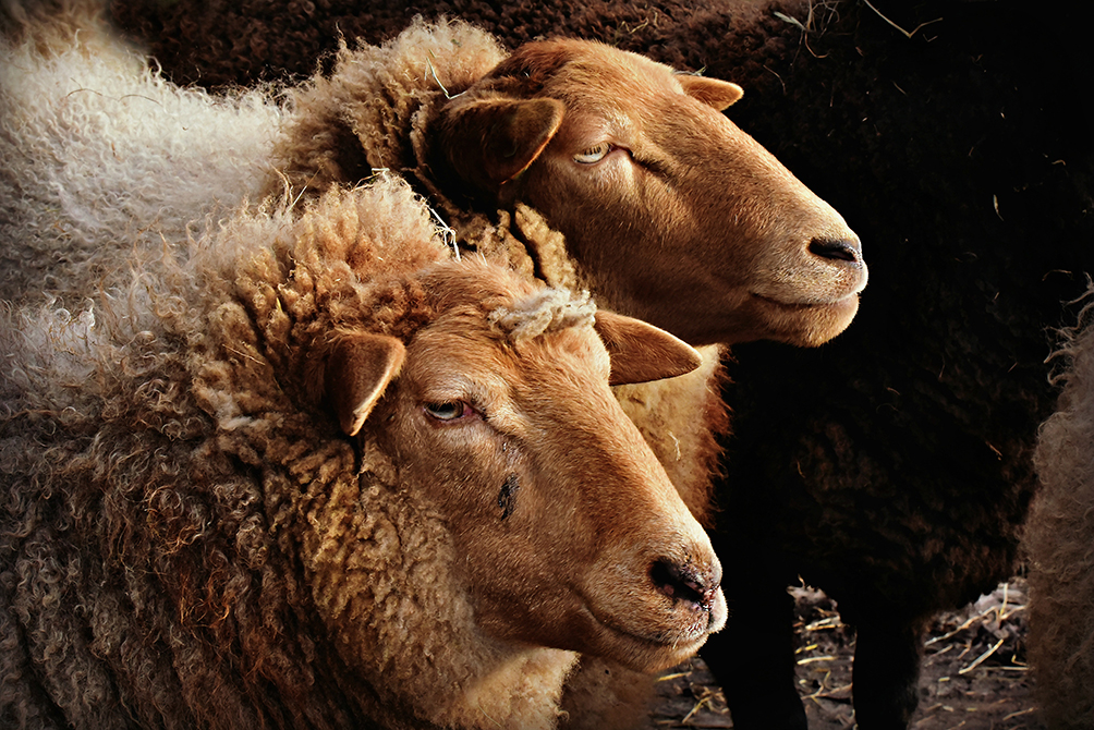
Although some areas of the country have been experiencing drought conditions, in other parts conditions appear to have been suitable for mud snails as we have been seeing evidence of fasciolosis in some of this year’s lambs for a few weeks now, which is quite early in the autumn for it. There have been cases in ewes and adult cattle too, but it is always possible that these could be due to infection last season.

Acorn poisoning

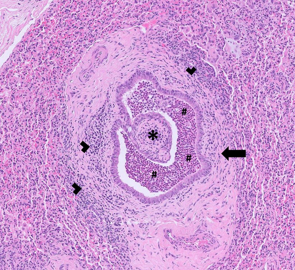
With some regions still suffering from a lack of grass the risk of acorn toxicity occurring is likely to be higher this year. Clinical signs can include dullness, anorexia, ruminal stasis, abdominal pain, tenesmus, constipation followed a few days later by dark diarrhoea, which can be mucoid and/or bloody. Subcutaneous oedema, which is often ventral, can also occur plus signs of renal failure, such as PUPD. Urea and creatinine levels are usually very high on blood testing due to severe renal tubular necrosis. Even if they survive the acute stage the prognosis is usually poor due to chronic renal failure. At PME there may be acorns or oak leaves in the rumen (if acute), haemorrhagic gastroenteritis, petechial haemorrhages, mucosal ulceration due to uraemia, ascites, hydrothorax and renal haemorrhages. Ocular fluid can be tested for urea and the histopathology findings in kidney can be pathognomonic for it. The area around oak trees should be fenced off, particularly if there is a shortage of grass.

New mastitis test

A new rapid, accurate and affordable molecular mastitis test is now available at Axiom. The test detects the presence of six key mastitis pathogens and the turnaround time is no longer than the following working day. This can allow a targeted course of treatment based upon the pathogens present, aiding responsible use of antibiotics and resulting in less discarded milk. As well as potentially saving money there are benefits for herd health in the long term. The agents detected are: E. coli, Staphylococcus aureus, Streptococcus uberis, Streptococcus dysgalactiae, Streptococcus agalactiae and Klebsiella pneumoniae. The cost of the test is £45 & VAT (test code FAMAS).

New more sensitive test for lungworm in cattle

We are pleased to be able to offer a PCR test for lungworm in cattle. As with the Baermann’s it detects the presence of larvae in the faeces (i.e. patent infections, from about 25 days post challenge). However, the PCR test does not require the larvae in faeces to be alive in order to obtain a positive result, which therefore increases the sensitivity. PCR tests are also usually more sensitive as they are able to detect very small amounts of an agent. Faecal samples should still be collected freshly voided or per rectum and kept cool until submission. Another benefit is that the test has been validated for the pooling of up to five samples, which makes it more cost effective to sample multiple animals. Ideally target animals that are coughing or showing suspicious respiratory signs. The turnaround time is the next working day, the test cost is £53 (& VAT) and the test code is PDVIV. We had three positive submissions with it in July – one of which was from adult beef cows.

Average test values (ATVs) for dairy herds testing using blood serology

For herds that are testing using bloods rather than milks for Johne’s serology an average test value can still be calculated but it is not directly comparable to the target used for milks. The Johne’s Action Group has advised that the ATV for bloods should be provided on the declaration form for the herd but state that it has been derived from blood testing. Dairy farmers using Johne’s blood serology can monitor the trend of their herd’s ATVs over time. We will automatically provide an ATV for herds that are testing through the Axiom Johne’s Monitoring Programme (plus any historic values since they started testing through the programme). If one is required when doing a 60 cow screen please request this on the submission form.

Animal Health & Welfare Pathway – worm egg counts now available

Please note that we are now participating in the worm egg counting part of the AHWP for sheep. However, we are unable to post out sampling kits. Consumables can be ordered from us in the usual way. WHEN SUBMITTING POST TREATMENT SAMPLES, PLEASE ENTER THE ACCESS (REPORT) NUMBER FOR THE PRE-TREATMENT SAMPLE RESULTS AS A PREVIOUS REFERENCE ON THE SUBMISSION FORM. We can then provide you with a % change in the strongyle egg count after treatment.
We are a UKAS accredited lab and provide ISO17025 accredited tests so we can carry out any of the follow up endemic disease testing for both cattle and sheep. The diseases and conditions to be sampled for sheep include: Border disease (BD), caseous lymphadenitis (CLA), enzootic abortion of ewes (EAE), Johne’s disease, Maedi Visna (MV), toxoplasmosis, tick-borne fever, pulpy kidney, lamb dysentery, ewe nutrition status, lamb nutrition status & trace elements. For cattle, a biosecurity assessment relating to BVD virus needs to be done in discussion with the farmer. This needs to cover whether or not it is appropriate for the herd to join a BVD accreditation programme. Membership is not compulsory, as many commercial herds may not be able to meet the rule requirements, particularly the requirement for a minimum three-metre biosecure gap. Vaccination also needs to be discussed – again it is not compulsory for it to be put in place though it is a good insurance policy until the country is further down the route of eradicating BVD virus. From the BVD check test results coming through the lab the vast majority of herds appear to be free of infection so could be becoming more and more naïve with time if they are not vaccinating. The impact of a BVD incursion, without the protection from BVD vaccination, could have a serious financial impact on a herd. Although a herd may be closed and appear to be well isolated we have seen breakdowns occurring due to the suspected transfer of virus on equipment or clothing.

BVD Cymru accredited lab

We are an accredited lab for the Welsh BVD eradication programme. BVD antibody and antigen results will be uploaded if samples are submitted on a BVD Cymru form. As was the case with BVD Free England, there is a small charge for the uploading of the results of 50p per sample for BVD antibody testing and 25p for a BVD antigen test. Please note that all fields on the BVD Cymru submission form must be completed (including the keeper’s phone number and email address) otherwise there is a block on the results uploading.

Discounted test rates for Johne’s disease & Neospora herd screens or regular batch testing

Our Johne’s & Neospora Monitoring programmes give farmers access to discounted test rates for whole herd or regular batch testing. There is no membership fee and no set rules to follow. Johne’s serology is from £5 per sample and Neospora serology costs from £6.75 per sample. It works out cheaper than testing through a CHECS cattle health scheme so is ideal for herds that are testing for disease control and management purposes. Advice is provided in the lab report and farmers can be e-mailed a copy if required. Our turnaround times are very fast– often same day but within three working days for both tests. Batch testing herds also get their results in a cumulative spreadsheet. A reminder to test email is sent out for herds on annual testing. Contact us for more information at dsfarm@axiomvetlab.co.uk or on 01626 357776.
In order to avoid any unnecessary confusion, please can we ask that submission forms are only sent in with the samples and not in advance of the samples. Thank you for your cooperation.

We would like your feedback on our services

As part of our continuing effort to help improve our services to clients within the Laboratory Division we have put together a short survey which will be sitting live permanently for anyone to complete whenever they have a few minutes spare. This can be filled in by anyone who uses any of our services and we encourage feedback to help us understand what we are doing well and where we need to make improvements. The QR code and link to the survey will remain live as a continuous tool to enable us to always gather feedback. If anyone has any questions about this they are welcome to contact either of the Quality Managers at the Laboratories, Claire Richardson for Axiom Veterinary Laboratories and Susan Reeve for Finn Pathologists. Thank you in advance for helping us to improve our services.

https://www.surveymonkey.com/r/Laboratory_Satisfaction_Survey
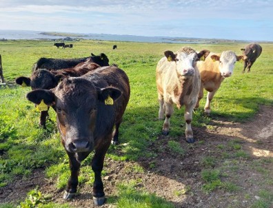
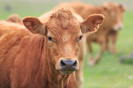
Interesting cases in July

Cattle
Abortions and mastitis

  • BVD virus was detected in foetal fluids of at least six abortion cases this month in the BVD antigen ELISA.
  • Neospora caninum was detected by PCR in at least four abortions including one of which was also positive for BVD antigen in foetal fluids and another from which Aerococcus sp. was cultured from foetal stomach contents. Aerococcus sp. is a common environmental bacterium but can act as an opportunistic pathogen causing sporadic abortions in cattle.
  • Coxiella burnetii (Q fever) was detected in placental tissue from at least three bovine abortions this month. In one of these cases the cow was reported to have aborted at seven months’ gestation and had retained placenta.
  • Salmonella species were detected by PCR in two cases of abortion this month. In one case culture was not possible and in the other the serotype was confirmed as Salmonella Dublin following culture. Salmonella sp. was isolated in selective cultures from the foetal stomach contents of an abortion which was the third to occur in a
    250-cow autumn block calving dairy herd. The herd had previously vaccinated but had stopped around two years previously. The serotype in this case was also confirmed as Salmonella Dublin.
  • Bacillus licheniformis was isolated from a five-month-gestation aborted bovine foetus. It was the third abortion to occur recently in the herd. All three aborted cows were well in themselves. Bacillus licheniformis is a known cause of abortion in cattle and may be associated with the ingestion of spoiled bedding or feed, or silage effluent.
  • Escherichia coli was isolated in pure growth from the stomach content of aborted foetus of a pedigree Hereford cow. Histopathology of liver tissue identified a multifocal subacute hepatitis consistent with systemic bacterial infection.
  • Trueperella pyogenes was isolated in two cases of abortion this month, one which was at five months’ gestation, the other at seven months.
  • Evidence of placentitis with elements of vasculitis and a corresponding bronchopneumonia suggestive of an infectious cause of abortion, most likely bacterial, was seen on histopathology of tissues from a seven-month gestation aborted foetus.
  • Of the less common pathogens isolated from milk samples this month, Pasteurella multocida was cultured from the milk of a three-year-old Holstein cow with episodes of mastitis recurring 5-10 days after a course of treatment producing very watery milk with large clots present. Nocardia sp. was also isolated from one sample.

Respiratory disease

  • Coronavirus was detected by PCR testing on nasal swabs in at least six submissions this month. In one case bought-in housed calves of around three months of age were showing signs of pyrexia and mortality in greater than two percent of the batch of 160. In another case there was evidence of clinical pneumonia in a group of 20 Wagyu calves which ranged from three to 12 weeks old. Coronavirus may be detected in nasal swabs from healthy animals as well as those showing signs of respiratory disease. All of the submissions which were positive on PCR for bovine coronavirus were also positive for one or more of the bacterial pathogens; Mycoplasma bovis, Pasteurella multocida, Mannheimia haemolytica or Histophilus somni. The finding of M. bovis, H. somni, P. multocida and M. haemolytica from nasal/NP swabs are of uncertain significance as they can be carried in the URT of healthy cattle. However, a study by Doyle (2017) found there was very good correlation between nasal or NP swabs or BAL samples and trans-tracheal washes with regards to M. haemolytica, Pasteurella multocida and Mycoplasma bovis. (They did not include H.somni in their study). (Doyle et al (2017) Agreement Among 4 Sampling Methods to Identify Respiratory Pathogens in Dairy Calves with Acute Bovine Respiratory Disease. J Vet Intern Med. 2017 May; 31(3):954-959).
  • PI3 and coronavirus were detected in one of three samples submitted from calves which were described as poor and scouring.
  • RSV was detected by PCR testing in at least two submissions. One case involved first season grazing heifers. Coughing and serous nasal discharge were seen in a significant proportion of the group of 200. The second case was in a ten-week-old calf purchased from market which had fever and open mouth breathing and later died. Consolidation and congestion were observed throughout the lung tissue at PME.
  • IBR was detected in a group of 12 to 16-month-old fattening bullocks in which six of the group of 20 had signs of pneumonia.
  • A fibrinous bronchopneumonia with pleurisy with appearance consistent with a case of ‘shipping fever’, caused by Mannheimia haemolytica was identified as the likely cause of death of an adult Holstein cow on histopathology. PCR testing of lung tissue was positive for both M. haemolytica and Pasteurella multocida. M. haemolytica was detected by PCR testing in lung tissue in at least two further cases of respiratory disease in adult dairy cattle. One of these was reported to have extensive lung consolidation at PME.
  • Pasteurella multocida, Mannheimia haemolytica and Histophilus somni were all detected by PCR in lung tissue from a Wagyu calf which was one of two to die from a recently purchased group of 40. A further 11 were affected with respiratory signs.
  • There were at least two submissions in which active seroconversion to Mycoplasma bovis was identified on paired serology following a respiratory disease outbreak in calves.
  • Bacterial bronchopneumonia consistent with descending infection with likely involvement of Pasteurellaceae was diagnosed by histopathology of lung tissue from a six-month-old dairy cross calf which was one of two to die in the group of 50 with a further four showing signs of weakness and scour.
  • Bronchointerstitial pneumonia was diagnosed as the likely cause of death in a four-month-old Limousin bull calf. The animal was found off-colour but deteriorated and died only 15 minutes later. Histopathology of lung tissue
    identified marked subacute broncho-interstitial pneumonia with hyaline membrane formation and early type II pneumocyte hyperplasia. A wide range of insults can result in interstitial pneumonia with hyaline membrane formation /alveolar epithelialisation in cattle including mouldy feedstuffs, pneumotropic viral infections and possibly idiosyncratic responses to 3methylindole (fog-fever like).
  • A one-day-old dairy heifer calf was euthanised on welfare grounds after showing signs of acute respiratory distress. Aspiration pneumonia was suspected to have occurred and this was supported by histopathology which identified severe inflammation and large amounts of protein rich fluid within the airways, consistent with milk/colostrum, likely administered inadvertently into the trachea by stomach tube.

  • Postmortem tissues were received from an adult Brown Swiss cow which was in a group of milking cows kept outdoors. Three cows had died. Ten percent were reported to have severe respiratory disease and a further 20 percent were coughing. Half the cows in the group had milk drop. IBR and RSV had been previously identified on the farm and this group had recently received an IBR vaccine. On histopathology there was a moderate-marked, multifocal-coalescing, subacute, eosinophilic and suppurative bronchointerstitial pneumonia with bronchiolitis fibrosa obliterans, intralesional nematode larvae and interlobular emphysema. The overall appearance with the presence of migrating larvae suggested a diagnosis of lungworm though it was not possible to rule out concurrent/previous viral insult/ inhaled pneumotoxin and/or secondary bacterial invasion. Lungworm was diagnosed by PCR testing of faeces in three cases and by Baermann’s examination in at least a further two cases this month. One case was in a group of four-month-old cross Jersey calves which had received ivermectin two weeks previous to sampling. Seroconversion to lungworm was seen in two cross Hereford female cattle with pyrexia and respiratory stertor.

Gastrointestinal disease

  • A total of 432 submissions for Johne’s disease serology and faecal PCR testing from cattle were received this month, with 132 submissions, including a total of 89 individual samples, testing positive. Blood and faecal samples were received from a cow which had a history of recent severe illness. It was the only animal in the group affected. Sudden weight loss and scour with haematochezia followed by dehydration and firm dung had been observed. Biochemistry revealed raised urea (43.7mmol/l: Reference interval 2.0-6.6mmol/l) and creatinine (448umol/l: Reference interval 40-170umol/l) indicating possible renal impairment. The animal tested strongly seropositive to Johne’s disease. Oak toxicity was considered a possible explanation for the azotaemia and acute signs of illness in this case. Johne’s disease was also diagnosed by serology in a two-year-old cow which had calved for the first time just five days previously but shown rapid loss in condition since. Another case was diagnosed on serology in a 12-month-old heifer, which was bought in four months previously and had been out at grass since, but was scouring and losing condition.
  • Coccidiosis was suspected following high counts in faecal samples along with consistent clinical history in numerous submissions this month. One case was in an 18-month-old heifer which had suddenly lost weight and had bloody diarrhoea. There was a coccidial oocyst of 18,300opg. On another holding a four-month-old Aberdeen Angus cross was found dehydrated with profuse bloody scour and had a coccidial oocyst count of >50,000opg, strongly suspicious of coccidiosis, though speciation was not carried out in that case. In a further submission Eimeria species identification was carried out on a faecal sample from a calf with acute illness and suspected coccidiosis despite a low coccidial oocyst count in a faecal egg count performed at the practice. 86% of the coccidial oocysts present were of the pathogenic species Eimeria bovis. In cases of coccidiosis, disease can precede patency so coccidiosis cannot be ruled out even with oocyst counts of <1000 opg.
  • Numerous cases of scour in calves due to Cryptosporidium were recorded. In two of these cases, hypogammaglobulinaemia was also identified indicating insufficient transfer of antibodies from colostrum. On one farm, calves which were reported to have inappetence and slight scour at 10-14 days of age tested positive for both Cryptosporidium and coronavirus on faecal sample testing. Diarrhoea caused by bovine coronavirus typically affects calves from three days to three weeks of age. Outbreaks may be similar or more severe than those caused by rotavirus. Affected calves will have liquid yellow diarrhoea which may contain mucus and blood. As with rotavirus infections, the clinical signs can be exacerbated by concurrent cryptosporidiosis.
  • Death due to E. coli F5 (K99) enteritis was suspected in at least two cases in neonatal calves this month.
  • There were at least six cases of disease associated with Salmonella Typhimurium, four of which were reported in freshly calved adult dairy cattle, one in young calves and there was one where no age was given. Salmonella Mbandaka was isolated from a four-year old, scouring, Holstein-Friesian cow on a farm where this serotype had been identified in the past. This serotype was also isolated from the faeces of a recently calved pyrexic dairy cow. Salmonella Dublin was isolated from two cases of scouring calves and another case where the age of animals was not stated. 
  • Yersinia enterocolitica and Yersinia pseudotuberculosis were isolated in faecal cultures from three to four-month-old calves which had been outdoors for one month and had watery/bloody diarrhoea and were rapidly losing condition. In-house coccidial oocyst counts had been very low. Subclinical carriage of Yersinia species is very common in young healthy ruminants. However, yersiniosis due to Yersinia pseudotuberculosis is a recognised cause of diarrhoea in calves often around four to ten months in age resulting in marked enteritis and formation of microabscesses within the intestinal mucosa. Conditions which may precipitate an overgrowth in the gut are trace element deficiency, parasitism, BVD infection, long transportation and malnutrition.
  • Raised pepsinogen indicating abomasal parasitism was recorded in numerous submissions this month. One case was a two-year-old Aberdeen Angus heifer which had deteriorated following calving and had a brown staring coat. The pepsinogen level was high at 5.79iu/l (RI <1.5iu/L). A poor doing diarrhoeic Holstein-Friesian heifer had a very high pepsinogen level of 7.25iu/L and was hypoalbuminaemic. Ostertagiosis was considered the likely cause of illness.

Skin and eye disease

  • Punch biopsies from masses on the skin of the flank of a Blonde d’Aquitaine cow were received for histopathology. The samples all comprised areas of ulcerated granulation tissue with prominent fibroblast proliferation, perivascular infiltrates of plasma cells and lymphocytes and multifocally accumulations of neutrophils surrounding collections of eosinophilic Splendore-Hoeppli material and accompanying epithelioid macrophages. The appearance was considered consistent with cutaneous bacterial infection with granulation, likely cutaneous actinobacillosis. This was further supported by gram staining which identified indistinct gram-negative rods associated with the centre of club colonies.
  • Histopathology of a biopsy from a mass on the penis of a 14-month-old Limousin bull identified the growth as a fibropapilloma. Fibropapillomas are hyperplastic lesions caused by papillomavirus infection. Penile fibropapillomas in bulls are most often diagnosed at 1-2 years of age. They can spontaneously resolve, but this occurs after a variable time period and recurrence can often be a problem. Rare cases can be persistent.
  • Moraxella bovoculi was isolated from two of four swabs submitted from a group of cattle in which an outbreak of conjunctivitis was spreading and which was poorly responsive to treatment.

Systemic and miscellaneous disease

  • A total of 378 submissions were received for BVD antigen testing in cattle in July, with 37 submissions, including a total of 50 individual samples, testing positive.
  • On one farm a seven-week-old calf with neurological signs was suspected to be a PI and was found to be BVD viraemic. The vet reported that there was no BVD control measures in place on the farm. On another farm a ‘bulldog’ calf tested positive for BVD virus in the antigen ELISA. On another holding a calf which was a suspected PI and had an irregular heart rhythm was confirmed as BVD viraemic in the antigen ELISA. A 10-month-old Belgian blue cross bullock which died after an acute illness tested positive for BVD virus by PCR on bloods with a low Ct value suggesting that the animal was likely to be a PI and the cause of death was suspected to be mucosal disease.
  • Antibodies to Border disease were detected in a bovine animal by SNT antibody testing to differentiate Border disease antibody from BVD antibody. The animal had repeatedly tested as suspect in the youngstock BVD antibody check test despite there being no evidence of a BVD breakdown in the herd. There were sheep present on the farm, but Border disease had not been previously identified in the flock.
  • Blood parasites consistent with Babesia spp. were seen on blood films from a cow in Devon with severe anaemia (PCV 12%) and jaundice. Biochemistry showed evidence of azotaemia most likely due to dehydration. The animal also had a high antibody titre to Anaplasma phagocytophilum (Tick borne fever) indicating recent exposure to this pathogen. Liver samples from a second cow from the same farm which had died showed peri-acinar hepatocellular necrosis and fatty change likely to be related to hypoxaemia associated with anaemia. On a farm in Cornwall, babesiosis was suspected and blood samples were submitted from the second cow to show clinical signs. Babesia sp. was detected by PCR in that case.
  • A 12-month-old bullock was found dead. The neck muscles were found to be oedematous, puffy and ‘crackly’ at PME. Acute necrotising and haemorrhagic myositis was identified on histopathology in the affected skeletal muscle and also in cardiac muscle. Gram-stained sections showed isolated slender gram-positive rod-shaped bacteria seen in association with areas of necrosis and haemorrhage consistent with a diagnosis of clostridial myositis (blackleg).
  • Two housed finishing heifers which died suddenly were found to have emphysematous lesions in the neck and shoulder muscles at PME. FAT testing of affected tissues from both animals was positive for Clostridium chauvoei and Clostridium septicum indicating evidence of both blackleg and pseudoblackleg in this case.
  • Two spring born dairy calves from a group of 300 were found dead. The herd had a history of deaths due to Histophilus somni and so the group had received an H. somni vaccine in March, though there was concern that immunological cover from this dose could now be waning. PME identified lesions suspicious of areas of necrosis in the heart but no significant findings in other major organ systems. Bacterial cultures of heart and lung tissue proved negative for significant pathogens, though Histophilus species are known to be fragile and may not survive long post-mortem. PCR testing was recommended and Histophilus somni was detected in heart muscle indicating this was the likely cause of the lesions and death in this case.
  • Nine cases of Malignant Catarrhal Fever were diagnosed by PCR testing. A PME was carried out on one of the cases and microscopic lesions consistent with MCF were seen (moderate, subacute, peri-portal lymphocytic and necrotising hepatitis with fibrinonecrotising vasculitis and mild to moderate peri-arteriolar lymphoid hyperplasia of the spleen).
  • Three male Aberdeen Angus cross cattle died with signs of uroabdomen and crystals around the preputial hairs following an issue with water availability which had occurred some weeks previously. Microscopy identified the crystals as struvite/ triple phosphate. Histopathology of kidney tissue identified severe chronic pyelonephritis with papillary necrosis. Water deprivation and reduced urinary flow resulting in ascending bacterial infection was considered to be the likely underlying pathogenesis in this case.
  • Severe fibrinous pleuritis and pericarditis with haemorrhage and acute interstitial pneumonia appeared to be the cause of death in a one-month-old Holstein-Friesian calf. The calf had been treated sporadically for pneumonia signs throughout its lifetime. Failure of transfer of passive immunity was suspected. Severe and locally extensive pleurisy with adhesions and haemorrhage were obvious at PME. On histopathology, the findings reflected acute fibrinous polyserositis with interstitial changes in the lung supporting a systemic inflammatory process/septicaemia. No evidence of primary bronchopneumonia was identified in this case.

Sheep and goats

Reproductive disease, mastitis

  • A very poor lambing season was experienced in a flock, with only one viable lamb from 12 ewes, lambs either being born dead or alive but struggling for air/gasping. All seven ewes sampled were seropositive for Toxoplasma.
  • Staphylococcus aureus was isolated from a chronic abscess on the udder of Charollais ewe and from a testicular abscess in an Oxford Down ram post vasectomy.

Respiratory disease

  • Several cases of suppurative, necrotising bacterial bronchopneumonia likely caused by members of the Pasteurellaceae were diagnosed on histopathology. In one case, a four-month-old lamb that died suddenly, there was also evidence of fibrinous pleuritis.
  • Post-mortem tissues were received from an older ewe, the latest of three ewes found dead with no preceding clinical signs. PME revealed multifocal lung adhesions and consolidated areas of lung with one lung lobe being firm and white in colour. Histopathology of lung was highly suggestive of ovine pulmonary adenocarcinoma due to Jaagsiekte retrovirus infection of sheep. Infection normally occurs at a young age with progressive neoplastic transformation of pulmonary tissue and the onset of clinical signs normally from two to three years of age. Animals commonly present with exercise intolerance and ill thrift however, if a secondary bacterial pneumonia develops, this can often lead to sudden death. In this case there was evidence of a secondary severe fulminating bacterial pneumonia which would have accounted for the sudden death in this animal.

Lung lobe effaced

  • A Kerry Hill ewe lamb was one of four to have died in a flock over the course of a week, and had been found struggling to breathe before dying, with profuse white foam emanating from the nostrils. On histopathology, the appearance was consistent with severe chronic bronchopneumonia. The presence of peribronchiolar fibrotic nodules accompanied by some hyperplasia were typical changes associated with ovine atypical pneumonia (Mycoplasma ovipneumoniae) infection with likely secondary bacterial involvement. M. ovipneumoniae is usually acquired in the lambing shed in the first few days of life and so attention to management factors at this stage (if lambed indoors) including improved ventilation and, if possible, early turnout of lambs can help mitigate problems associated with this pathogen. Severity of pathology associated with this infection can vary and clinical consequences vary from reduced live weight gain to severe pneumonia and death, usually associated with secondary bacterial infection. Recent herding and introduction of new animals into the flock can also be associated with outbreaks of atypical pneumonia.

Gastrointestinal disease

  • Among the cases of coccidiosis was a two to three-month-old lamb with an in house coccidial oocyst count of 277,800 opg of which approx. two thirds of the oocysts were identified as the pathogenic Eimeria species, E. ovinoidalis and E. crandallis.
  • Several cases of parasitic gastroenteritis were diagnosed in sheep flocks and goat herds, with signs including weight loss, scour and acute deaths. A number of cases of haemonchosis were suspected, with high strongyle egg counts and signs including submandibular oedema, anaemia and weight loss. Some cases were confirmed on peanut agglutinin testing.
  • Fasciolosis was diagnosed on microscopy and liver fluke coproantigen ELISA in some flocks this month. In one flock, a ewe in poor condition and with scour was diagnosed with fasciolosis and Johne’s disease on faecal PCR. Fasciolosis was also diagnosed on histopathology of liver of a ewe in another flock.

A fluke in the liver

  • Post-mortem tissues were received from an 18-month-old female Golden Guernsey goat. The goat had presented clinically with opisthotonus, circular nystagmus, an unreliable menace response and pyrexia (39.6oC). Despite treatment, the animal died overnight, with the main PME finding being toxic looking small intestines. Clostridium perfringens alpha and epsilon toxins were detected on ELISA in small intestinal contents and there was also perivascular oedema in the brain providing further support for a diagnosis of Clostridium perfringens type D enterotoxaemia. The goat was not vaccinated against clostridial disease. This was also suspected to be the cause of sudden death in a four-month-old Jacob lamb that died acutely. Again, C. perfringens alpha and epsilon toxins were detected in small intestinal contents and the lamb had not received its full primary course of clostridial vaccination.
  • A total of 96 submissions for Johne’s disease serology and faecal PCR testing from sheep and goats were received this month, with 11 submissions, including a total of 17 individual samples, testing positive. Cases included a Dorset tup that was losing body condition (BCS 2/5) and was lethargic and pyrexic on clinical examination, and a Valais Blacknose wether that was chronically wasted and scouring.

Skin and eye disease

  • Actinobacillus sp. was isolated from an abscess on the neck of a tup, consistent with a diagnosis of cutaneous actinobacillosis. Skin trauma is thought to allow entry of bacteria into the skin.
  • A Bovicola sp. (biting louse) infestation was confirmed on skin microscopy in a goat with hair loss over the back and ears that was only partially responsive to ivermectin treatment.
  • Seroconversion to sheep scab was seen in some flocks with a history of pruritus, wool loss and/or scabs/scaling of skin.
  • Sections of eyelid and ear were submitted from a sheep with multiple skin masses. Histopathology was consistent with destructive and highly infiltrative squamous cell carcinoma. In sheep, this is most common on the muzzle, lips, eyelids, ears, perineum and vulva. There is an increased incidence in Merino sheep and exposure to UV light may play a role in development. In general, squamous cell carcinomas are locally invasive neoplasms with a relatively low rate of metastatic spread, which may occur later in the course of the disease. Visceral metastasis from a cutaneous squamous cell carcinoma has been recorded in sheep.

Systemic and miscellaneous disease

  • Two sheep were confirmed as persistently infected with Border disease using the BVD antigen ELISA test which we have validated for detection of BDV PIs. In an Easycare flock, six out of twelve 15-month-old ewe lambs tested were seropositive for BDV, consistent with exposure to the virus.
  • A thin pygmy goat that had lost a moderate amount of body condition despite eating satisfactorily and became lame on the right fore was seropositive to CAE.
  • Several flocks had seropositive animals when screening for Maedi visna. Where clinical history was provided, ill thrift was the primary presenting sign.
  • A group of lambs were brought back in off the hill after becoming ill thrifty, pyrexic, lethargic and developing swollen joints. Anaplasma phagocytophilum was detected by PCR in EDTA blood samples from two animals confirming Tick Borne Fever.
  • Two cases of cerebrocortical necrosis/polioencephalomalacia were diagnosed. Typically, cases are due to thiamine deficiency or sulphur toxicity. The former is commonly associated with recent changes in diet resulting in rumen dysbiosis and the production of thiaminase with subsequent vitamin B1 deficiency. Sulphur toxicity is associated with excess intake through concentrate food and/or water. One of the two sheep also had lung histopathology consistent with atypical/epizootic pneumonia.
  • A blood sample from a four-month-old Hampshire cross ewe lamb detected a very high urea level of 177.8 mmol/l (RI: 2.6-6.6) likely to be consistent with nephrosis. The precise aetiology of the condition is unknown, but it may occur in association with nematodirosis and coccidiosis. A relatively low coccidial oocyst count of 1950 opg was present but this was not speciated.
  • A two-year-old Texel wether developed a small, 50p sized lump on the thorax last year and, by May, this had grown to an approx. 12 x 14cm, weeping, fissured lesion in the skin which the animal was self-traumatizing. Skin nodules were present which appeared to grow in number after debridement, were white to cream in colour, soft and malleable, appearing to be fluid-filled and ranging in size from 3 x 3 mm to 1 x 1 cm. Histopathology of submitted biopsies revealed changes consistent with a round cell tumour, the most likely differentials including a mast cell tumour and lymphoma. No metachromatic granules were seen with Giemsa special stain to suggest a mast cell proliferation and therefore it was suspected to be a lymphoma.
    Round cell tumour

Deficiencies and toxicities

  • There were several diagnoses of cobalt deficiency based on low vitamin B12 levels in serum in sheep this month, two in goats and two in camelids. Where history was available in the sheep flocks the main clinical sign was poor body condition. One affected sheep flock reported recurrent skin issues with lambs developing crusting of the ear pinnae during the summer. Other submissions reported concurrent heavy parasite burdens or suspected haemonchosis and a further flock had a history of issues with mastitis in ewes.
  • Copper deficiency was suspected to be involved in problems in second calvers with fertility issues, poor body condition and diarrhoea. Three cows were tested from the group and all three had low blood copper. There was
    no evidence of seroconversion to Fasciola hepatica or Johne’s disease on serology. A group of Simmental cows were reported to be losing condition and some had gone down after calving. All five animals tested were hypocupraemic. In another suckler herd, which reported that calves were weak this year, all five cows tested had very low copper levels with the mean for the group being 3.2umol/l (RI 9.0-19.0umol/l). Plasma inorganic iodine was also low for the group at 39ug/l (levels >150ug/l for a group indicates adequate intake) indicating possible iodine deficiency as well as copper deficiency. Three 10-week-old goat kids in a group of five showed signs of progressively weakening hindlimbs and ataxia. All five kids had very low copper levels with a mean copper level for the group of 1.2umol/l (RI 9.0 -25.0umol/l). The findings were suspicious of a diagnosis of delayed swayback (enzootic ataxia) caused by copper deficiency.
  • Copper toxicity was suspected in a cow which died after a short illness lasting around 24 hours. Haematuria and sudden milk drop were observed and the animal died after developing ruminal bloat. At PME, it was noted that the liver was orange in colour. Biochemistry of blood samples taken premortem showed severely raised liver enzymes with a GLDH level of 6408 U/L (RI 0-25 U/L) and GGT 828 U/L (RI 0-50 U/L). Histopathology of liver tissue identified severe peri-acinar hepatocellular necrosis. Special staining did not identify any evidence of copper accumulation in liver or kidney tissue. Other possible causes of severe hepatocellular necrosis such as blue-green algae toxicity were considered. Liver tissue copper was high at 11522umol/kg DM (RI 314.00-10000umol/kg) and in the light of this finding copper toxicity was considered to be the most likely explanation for the death despite negative staining on histopathology.
  • Hypocalcaemia was identified in at least 13 cases this month, twice in combination with hypophosphataemia and in one animal which was also hypomagnesaemic. All the cows were reported to be down cows where history was available. One animal was reported to also have retained foetal membranes and metritis, one had a leg abscess and another cow had suspected ragwort toxicity. One case of hypomagnesaemia tested positive for Salmonella Mbandaka on faecal cultures. The concurrent conditions were likely to have predisposed these cows to hypocalcaemia/ hypomagnesaemia.
  • Ketosis was identified in a down Jersey cow that appeared to have an ‘S’ bend in the neck indicative of milk fever, though there was no response to calcium administration. BHB was raised at 3.46 mmol/l (RI: 0.0-1.2 mmol/l) and NEFA levels were also high at 1439 μmol/l (RI 0-600 μmol/l) suggesting excessive mobilisation of fat to meet energy needs and a high risk of developing fatty liver disease. GLDH was moderately raised at 242 U/L (RI 0-25 U/L) and GGT mildly raised at 99 U/L (RI 0-50 U/L) raising the suspicion that fatty infiltration of the liver could be occurring.
  • Low plasma inorganic iodine levels in pooled samples indicating low dietary iodine intake were thought to be the possible cause of poor scanning results in one group of cows and a group of ewes from different farms.
  • A high plasma inorganic iodine level was recorded in a pooled sample from one farm experiencing premature calvings, stillbirths and calves which were slow to stand following birth. PII reflects only recent dietary intake so even a high result cannot rule out deficiency during earlier gestation, particularly if there has been a change in diet. Excess iodine in the diet has been associated with poor passive transfer of IgG in sheep. Iodine toxicity can result in abortions in cattle.
  • Selenium and cobalt toxicity were suspected in a group of cheviot lambs following an accidental 10x overdose of a trace element drench. Eighteen animals from the group of 40 died overnight. Postmortem findings were unremarkable. Liver from two animals was tested for trace elements and selenium and cobalt were at toxic levels in both animals.
  • White muscle disease due to selenium deficiency was suspected to be the cause of sudden death in a six-week-old suckler calf. Very pale muscles were observed at PME. The liver selenium level was indicative of deficiency at 3.67 μmol/kg DM (RI 11.0-22.6 μmol/kg DM). Copper and manganese were marginal. Histopathology identified basophilic granular material within Purkinje cells of the heart, which Von Kossa staining revealed to be mineral deposits. Mineralisation of Purkinje cells has been previously seen in calves with selenium/ vitamin E deficiency and, although not diagnostic of white muscle disease, the finding provided supportive evidence in this case. It is thought that mineralisation within the Purkinje cardiocytes can interfere with the cardiac conduction system and this could have been responsible for the sudden death in this case.

Lakes of dense basophilic fluid

  • Vitamin A deficiency was thought be the possible cause of blindness in seven-week-old twin calves. Both had vitamin A levels below the expected reference range at 163 μg/l and 153 μg/l (RI 249-500 μg/l). Persistent BVD infection was considered as a possible differential but both calves tested negative in the BVD antigen ELISA.
  • There was evidence of zinc deficiency and low plasma inorganic iodine in animals tested from a group of cows with a history of poor growth rates in their calves.

Camelids

  • A one-month-old alpaca cria with abdominal pain but normal pelleted faeces had a coccidial oocyst count of greater than 50,000 opg. Although no Eimeria macusaniensis oocysts were seen, the smaller Eimeria species in camelids are reported to be pathogenic in crias, suggesting this was a case of coccidiosis.
  • Several cases of parasitic gastroenteritis were reported in alpacas with clinical signs including weight loss and scour. In one case, a strongyle egg count of 6950 epg, potentially consistent with haemonchosis, was obtained from a faeces sample from a male alpaca 10 days after ivermectin treatment, suggestive of treatment failure although a pre-treatment strongyle egg count had not been obtained. PGE was also diagnosed in a scouring, ill thrifty camel.
  • A three-year-old female alpaca developed weight loss, inappetence, respiratory compromise and generalised lymphadenopathy four weeks post-partum, with minimal response to antibiotic treatment. Evaluation of a blood smear revealed markedly elevated numbers of lymphoid cells (117 x 109/l) comprising a mixture of approximately equal numbers of well-differentiated small lymphocytes and similar appearing, intermediate lymphoid cells with slightly more abundant, mid basophilic cytoplasm, which occasionally had peripheral blebs, and an eccentric round or irregular round nucleus with coarse chromatin and sometimes an indistinct nucleolus. The findings were consistent with chronic lymphocytic leukaemia. Aspirates taken from one prescapular lymph node only detected reactive lymphoid tissue with mild neutrophilic inflammation.
  • After a previous positive result in an alpaca herd, seven animals were screened for Johne’s disease on faecal PCR and all seven were positive. No further history was supplied.
  • A 12-month-old male llama died after a 24-hour history of lethargy. On PME, the small intestine appeared thickened and there were white plaques on the distal trachea. Histopathology detected an acute necrotising enteritis and a bacterial aetiology appeared likely. There was also a mild multifocal bronchopneumonia associated with fungal hyphae which may have represented aspirated material from the fungal plaques seen on the trachea.

Other Species

  • Fasciolosis was diagnosed on routine monitoring coproantigen ELISA testing in an adult reindeer.
  • Mycoplasma gallisepticum infection was confirmed on PCR testing of an ocular swab from three to four-week-old pheasant chicks.
  • A mass on the foreleg of a euthanased fallow deer was found to be a soft tissue sarcoma on histopathology, potentially a fibrosarcoma.
  • A moderate, pure growth of E. coli was isolated from a yolk sac infection in a five-day-old pheasant chick.

Follow us on Facebook

https://www.facebook.com/Axiomfarmvetlab/

This newsletter is sent by e-mail to each vet practice but if you would like a copy sent to your individual e-mail account please contact us at dsfarm@axiomvetlab.co.uk and we can add you to our circulation list.

Related Posts