Email Us
admin@axiomvetlab.co.uk

admin@axiomvetlab.co.uk
(01626) 355655

This newsletter is sent by e-mail to each vet practice but if you would like a copy sent to your individual e-mail account please contact us at dsfarm@axiomvetlab.co.uk and we can add you to our circulation list.
We are pleased to be able to offer a PCR test for lungworm in cattle. As with the Baermann’s it detects the presence of larvae in the faeces (i.e. patent infections, from about 25 days post challenge). However, the PCR test does not require the larvae in faeces to be alive in order to obtain a positive result, which therefore increases the sensitivity. PCR tests are also usually more sensitive as they are able to detect very small amounts of an agent. Faecal samples should still be collected freshly voided or per rectum and kept cool until submission. Another benefit is that the test has been validated for the pooling of up to five samples, which makes it more cost effective to sample multiple animals. Ideally target animals that are coughing or showing suspicious respiratory signs. The turnaround time is the next working day, the test cost is £53 (& VAT) and the test code is PDVIV.
For many tests, sending samples with an ice pack particularly in times of hot weather is a good idea to ensure the sample arrives with us in the best possible condition. If you are planning to do this, please can you make sure that the ice pack is placed inside the UN3373 courier bag packaging.
For herds that are testing using bloods rather than milks for Johne’s serology an average test value can still be calculated but it is not directly comparable to the target used for milks. The Johne’s Action Group has advised that the ATV for bloods should be provided on the declaration form for the herd but state that it has been derived from blood testing. Dairy farmers using Johne’s blood serology can monitor the trend of their herd’s ATVs over time. We will automatically provide an ATV for herds that are testing through the Axiom Johne’s Monitoring Programme (plus any historic values since they started testing through the programme). If one is required when doing a 60 cow screen please request this on the submission form.
As part of our continuing effort to help improve our services to clients within the Laboratory Division we have put together a short survey which will be sitting live permanently for anyone to complete whenever they have a few minutes spare. This can be filled in by anyone who uses any of our services and we encourage feedback to help us understand what we are doing well and where we need to make improvements. The QR code and link to the survey will remain live as a continuous tool to enable us to always gather feedback. If anyone has any questions about this they are welcome to contact either of the Quality Managers at the Laboratories, Claire Richardson for Axiom Veterinary Laboratories and Susan Reeve for Finn Pathologists. Thank you in advance for helping us to improve our services.
https://www.surveymonkey.com/r/Laboratory_Satisfaction_Survey

When sending submissions containing 50+ bloods, please ensure that you use a Field Kit containing a polystyrene sample rack/box for the orderly transportation of your samples, remembering to populate the rack in the same order as your accompanying animal ID list. Receiving large quantities of blood samples in a plastic bag or cardboard box is not appropriate or conducive to the efficient handling of such submissions and invariably leads to significant delays in preparation and turnaround times. Field Kits (filled with the required serum gel tubes) are readily available to order via the following link…https://milab.store.unleashedsoftware.com/
Please note that we are now participating in the worm egg counting part of the AHWP for sheep. However, we are unable to post out sampling kits. Consumables can be ordered from us in the usual way. WHEN SUBMITTING POST TREATMENT SAMPLES, PLEASE ENTER THE ACCESS (REPORT) NUMBER FOR THE PRE-TREATMENT SAMPLE RESULTS AS A PREVIOUS REFERENCE ON THE SUBMISSION FORM. We can then provide you with a % change in the strongyle egg count after treatment.
We are a UKAS accredited lab and provide ISO17025 accredited tests so we can carry out any of the follow up endemic disease testing for both cattle and sheep.
The diseases and conditions to be sampled for sheep include:
Border disease (BD), caseous lymphadenitis (CLA), enzootic abortion of ewes (EAE), Johne’s disease, Maedi Visna (MV), toxoplasmosis, tick-borne fever, pulpy kidney, lamb dysentery, ewe nutrition status, lamb nutrition status & trace elements. For cattle, a biosecurity assessment relating to BVD virus needs to be done in discussion with the farmer. This needs to cover whether or not it is appropriate for the herd to join a BVD accreditation programme. Membership is not compulsory, as many commercial herds may not be able to meet the rule requirements, particularly the requirement for a minimum three-metre biosecure gap. Vaccination also needs to be discussed – again it is not compulsory for it to be put in place though it is a good insurance policy until the country is further down the route of eradicating BVD virus. From the BVD check test results coming through the lab the vast majority of herds appear to be free of infection so could be becoming more and more naïve with time if they are not vaccinating. The impact of a BVD incursion, without the protection from BVD vaccination, could have a serious financial impact on a herd. Although a herd may be closed and appear to be well isolated we have seen breakdowns occurring due to the suspected transfer of virus on equipment or clothing.
Please provide your name, practice, the farmer and farm names so that we can link the photos to the relevant submission and please also indicate which Axiom vet you discussed the case with. We may wish to use some of the photos in our newsletter so please indicate if you are not happy for this to be done. All cases are anonymised and credited to the submitting vet. Please note that this number is just for sharing photos. If you wish to discuss a case for which you do not have photos, please ring 01626 357776 as usual.
We are an accredited lab for the Welsh BVD eradication programme. BVD antibody and antigen results will be uploaded if samples are submitted on a BVD Cymru form. As was the case with BVD Free England, there is a small charge for the uploading of the results of 50p per sample for BVD antibody testing and 25p for a BVD antigen test.
Please note that all fields on the BVD Cymru submission form must be completed (including the keeper’s phone number and email address) otherwise there is a block on the results uploading.
Our Johne’s & Neospora Monitoring programmes give farmers access to discounted test rates for whole herd or regular batch testing. There is no membership fee and no set rules to follow. Johne’s serology is from £4.25 per sample and Neospora serology costs from £5.90 per sample. It works out cheaper than testing through a CHECS cattle health scheme so is ideal for herds that are testing for disease control and management purposes. Advice is provided in the lab report and farmers can be e-mailed a copy if required. Our turnaround times are very fast– often same day but within three working days for both tests. Batch testing herds also get their results in a cumulative spreadsheet. Contact us for more information at dsfarm@axiomvetlab.co.uk or on 01626 357776.
In order to avoid any unnecessary confusion, please can we ask that submission forms are only sent in with the samples and not in advance of the samples. Thank you for your cooperation.
You may find this is a more efficient way of making requests than phoning the farm team, saving you time in your busy day. Our farm team also find it a more efficient way of dealing with your requests.
The email address for test requests is: DSFarm@axiomvetlab.co.uk
Cattle
Abortions and mastitis
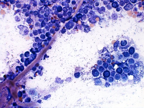
Respiratory disease
Abundant degenerate neutrophils (often with streaming nuclei; oat cells) (arrows) and fibrin (*) in a calf with fulminating bacterial pneumonia likely caused by Mannheimia haemolytica
Gastrointestinal disease
Skin and eye disease
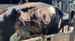
Severe necrotising dermatitis in a British Friesian cow (photo: Eleanor Stafford, Axe Valley Large Animal Vets)
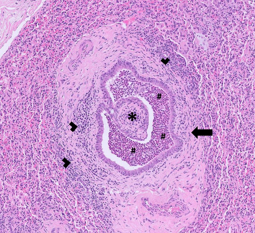
Systemic and miscellaneous disease
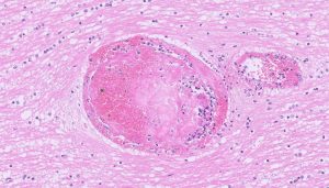
Vasculitis and thromboembolism of a vessel in the brain of a fattening heifer with histophilosis
Abortion, infertility and mastitis
Respiratory disease
Gastrointestinal disease
Skin and eye disease
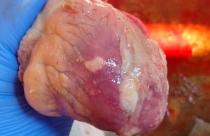
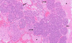
Pale lesions consistent with myocarditis in a lamb with tick pyaemia (Photo: Jess Kebbell, Derwent Vale Farm Vets)
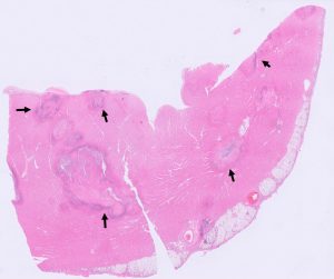
Multifocal areas of suppurative myocarditis (arrows) in the heart of a lamb with tick pyaemia
Farm May Newsletter 2026 Dedicated farm line – 01626 357776 Follow us on Facebook https://www.facebook.com/Axiomfarmvetlab/…
Farm April Newsletter 2026 Dedicated farm line – 01626 357776 Follow us on Facebook https://www.facebook.com/Axiomfarmvetlab/…
Farm March Newsletter 2026 Dedicated farm line – 01626 357776 Follow us on Facebook https://www.facebook.com/Axiomfarmvetlab/…
Download a PDF
The great explorer of the truth, the master-builder of human happiness no one rejects dislikes avoids pleasure itself because it is pleasure but because know who do not those how to pursue pleasures rationally encounter consequences that are extremely painful desires to obtain.
Read More