Farm April Newsletter 2025

Dedicated farm line – 01626 357776

This newsletter is sent by e-mail to each vet practice but if you would like a copy sent to your individual e-mail account please contact us at dsfarm@axiomvetlab.co.uk and we can add you to our circulation list.


We would like your feedback

As part of our continuing effort to help improve our services to clients within the Laboratory Division we have put together a short survey which will be sitting live permanently for anyone to complete whenever they have a few minutes spare. This can be filled in by anyone who uses any of our services and we encourage feedback to help us understand what we are doing well and where we need to make improvements. The QR code and link to the survey will remain live as a continuous tool to enable us to always gather feedback. If anyone has any questions about this they are welcome to contact either of the Quality Managers at the Laboratories, Claire Richardson for Axiom Veterinary Laboratories and Susan Reeve for Finn Pathologists. Thank you in advance for helping us to improve our services.

https://www.surveymonkey.com/r/Laboratory_Satisfaction_Survey


Please use field kits with herd screens

When sending submissions containing 50+ bloods, please ensure that you use a Field Kit containing a polystyrene sample rack/box for the orderly transportation of your samples, remembering to populate the rack in the same order as your accompanying animal ID list. Receiving large quantities of blood samples in a plastic bag or cardboard box is not appropriate or conducive to the efficient handling of such submissions and invariably leads to significant delays in preparation and turnaround times.
Field Kits (filled with the required serum gel tubes) are readily available to order via the following link… https://milab.store.unleashedsoftware.com/

Diagnosing Winter Dysentery

We have two testing options for winter dysentery – paired coronavirus serology to check for rising antibody levels or PCR testing for the presence of virus using faecal samples. The vast majority of adult cattle have detectable antibodies (though immunity does not appear to be complete) therefore in order to demonstrate recent exposure to coronavirus it requires paired serology to check for rising antibody levels. Blood samples should be collected 2-4 weeks apart. Alternatively, for a quicker result, faeces can be tested for the virus using a PCR test. Virus tends to only
be shed for a very short time following infection so it is important that new cases are sampled. Although the PCR test has not been validated for testing of pooled samples we seem to have had better success with detecting it in pooled rather than individual faecal samples. With it being a PCR test it is a sensitive test and by sampling multiple animals it increases the chance of detecting the virus. We would recommend no more than three faecal samples in a pool to reduce the risk of over dilution with negative samples, which could compromise sensitivity.

Animal Health & Welfare Pathway – worm egg counts now available

Please note that we are now participating in the worm egg counting part of the AHWP for sheep. However, we are unable to post out sampling kits. Consumables can be ordered from us in the usual way. WHEN SUBMITTING POST TREATMENT SAMPLES, PLEASE ENTER THE ACCESS (REPORT) NUMBER FOR THE PRE-TREATMENT SAMPLE RESULTS AS A PREVIOUS REFERENCE ON THE SUBMISSION FORM. We can then provide you with a % change in the strongyle egg count after treatment.
We are a UKAS accredited lab and provide ISO17025 accredited tests so we can carry out any of the follow up endemic disease testing for both cattle and sheep. The diseases and conditions to be sampled for sheep include: Border disease (BD), caseous lymphadenitis (CLA), enzootic abortion of ewes (EAE), Johne’s disease, Maedi Visna (MV), toxoplasmosis, tick-borne fever, pulpy kidney, lamb dysentery, ewe nutrition status, lamb nutrition status & trace elements.

Would you like to send the Axiom farm vets photos of PMEs to allow us to assist with sample selection and diagnosis? Please send photos by WhatsApp to 07944 649222.

Please provide your name, practice, the farmer and farm names so that we can link the photos to the relevant submission and please also indicate which Axiom vet you discussed the case with. We may wish to use some of the photos in our newsletter so please indicate if you are not happy for this to be done. All cases are anonymised and credited to the submitting vet. Please note that this number is just for sharing photos. If you wish to discuss a case for which you do not have photos, please ring 01626 357776 as usual.

BVD Cymru accredited lab

We are an accredited lab for the Welsh BVD eradication programme. BVD antibody and antigen results will be uploaded if samples are submitted on a BVD Cymru form. As was the case with BVD Free England, there is a small charge for the uploading of the results of 50p per sample for BVD antibody testing and 25p for a BVD antigen test.
Please note that all fields on the BVD Cymru submission form must be completed (including the keeper’s phone number and email address) otherwise there is a block on the results uploading.

Discounted test rates for Johne’s disease & Neospora herd screens or regular batch testing

Our Johne’s & Neospora Monitoring programmes give farmers access to discounted test rates for whole herd or regular batch testing. There is no membership fee and no set rules to follow. Johne’s serology is from £4.25 per sample and Neospora serology costs from £5.90 per sample. It works out cheaper than testing through a CHECS cattle health scheme so is ideal for herds that are testing for disease control and management purposes. Advice is provided in the lab report and farmers can be e-mailed a copy if required. Our turnaround times are very fast– often same day but within three working days for both tests. Batch testing herds also get their results in a cumulative spreadsheet. Contact us for more information at dsfarm@axiomvetlab.co.uk or on 01626 357776.
In order to avoid any unnecessary confusion, please can we ask that submission forms are only sent in with the samples and not in advance of the samples. Thank you for your cooperation.

Did you know that you can email us any additional test requests for your submissions?

You may find this is a more efficient way of making requests than phoning the farm team, saving you time in your busy day. Our farm team also find it a more efficient way of dealing with your requests.
The email address for test requests is: DSFarm@axiomvetlab.co.uk
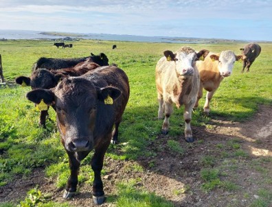
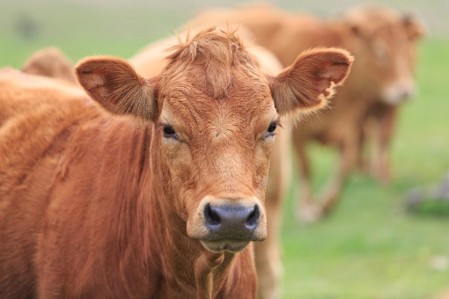
Interesting cases in February

Cattle
Abortions and mastitis

  • Bacillus licheniformis was cultured from two bovine abortion submissions this month, both of which occurred in late gestation.
  • Two Hereford cows in a 40-cow herd both aborted on the same day around the eighth month of pregnancy. Listeria monocytogenes was cultured from one of the two foetuses. In another Listeria abortion, case a Friesian cow aborted at eight months gestation and was the fifth in the herd to abort recently. Listeria monocytogenes was identified by both culture and PCR testing in that case.
  • Salmonella Mbandaka was identified as the isolate in a case of abortion at six months gestation.
  • Staphylococcus vitulinis was isolated in moderate growths from an eight months gestation aborted foetus out of a Charolais heifer. The isolate in this case was considered an unlikely pathogen but it couldn’t be ruled out as a possible cause of sporadic abortion.
  • Neospora caninum was detected by PCR in foetal tissues of a cow that aborted in the sixth month of pregnancy and was otherwise clinically well.
  • Antibodies to Neospora caninum were detected in the foetal fluids of stillborn calves in two submissions. In one case, the dam was reported to have aborted the previous year.
  • Leptospira sp. was detected by PCR of foetal kidney in one case of bovine abortion this month.
  • Pasteurella multocida was isolated from a milk sample from a two-year-old lactating heifer with chronic recurring mastitis that had been treated with multiple antibiotics. P. multocida is an uncommon cause of mastitis in cows and in some case can be fatal due to the presence of endotoxins.
  • Candida sp. mastitis was identified in two cows on the same farm with clinical mastitis and the presence of clots.

Respiratory disease

  • IBR virus was detected in several respiratory cases this month. In one case, an animal with purulent nasal discharge died and purulent material was observed in the trachea at PME. PCR testing of lung tissue detected IBR along with Mycoplasma bovis, Histophilus somni and Pasteurella multocida. Lung histopathology was supportive of the involvement of all these respiratory pathogens (although the bacteria are likely to be opportunistic invaders).
  • RSV was detected as the primary respiratory pathogen by PCR testing in at least 36 submissions, in combination with PI3 in at least four submissions, in combination with Bovine Coronavirus in at least 16 submissions and with both PI3 and Coronavirus in at least one case. In a number of cases, bacterial respiratory pathogens were also detected and where these were detected in lung tissue or the lower airways these were considered significant (at least as opportunistic pathogens). RSV was detected in lung tissue from a three-month-old calf that was the only mortality in a group of 50 that had sudden onset coughing and pyrexia. Abscessation was observed in the lung tissue at PME and histopathology confirmed chronic-active, plasmalymphocytic and suppurative bronchointerstitial pneumonia with bronchiolitis fibrosa obliterans consistent with viral infection and secondary bacterial infection. Histopathology remains the best single method of characterising the complexity of bovine respiratory disease, when a carcase is available.
  • PI3 was detected by PCR in at least five further cases of bovine respiratory disease this month and in combination with coronavirus in at least three cases.
  • Mannheimia haemolytica was isolated in cultures of lung tissue from an eight-week-old weaned Holstein-Friesian dairy calf that died after a short bout of respiratory illness. A marked fibrinous reaction was observed at PME throughout the thorax and abdomen. Histopathology identified a fibrinosuppurative pleuritis consistent with Mannheimia infection and the presence of fibrin in the abdominal cavity (polyserositis) suggested that the lung changes were part of a septicaemic process rather than primary pneumonia. Underlying risk factors can include failure of passive transfer, underlying disease (e.g. BVD), trace element deficiencies, and stress due to moving, mixing or adverse weather.
  • Pasteurella multocida was identified by PCR testing of lung tissue from youngstock with respiratory disease. In one case, histopathology identified a likely primary viral insult with evidence of secondary infection.
  • A seven month-old heifer recently purchased from market along with four others from a different farm was found down in the shed in respiratory distress and unable to rise. It had been noted by the farmer that there had been some coughing in the group. PCR’s of affected lung tissue detected Mycoplasma bovis, Pasteurella multocida and Mannheimia haemolytica. There were severe lung changes seen on histopathology. A severe, extensive, chronic-active, necrosuppurative and fibrinohaemorrhagic bronchointerstitial pneumonia with intralesional bacteria was confirmed .The predominant lesions were an acute alveolar necrosis (which can be caused by either Mycoplasma bovis or Mannheimia haemolytica), acute suppurative infiltrate (associated with M. haemolytica) and caseonecrotic airways (caused by M. bovis). Co-infections with M. bovis and M. haemolytica are common, and recent studies suggest M. haemolytica results in worsening of the respiratory disease caused by M. bovis.
  • Mycoplasma bovis, Pasteurella multocida and Histophilus somni were detected by PCR of lung tissue from a four-month-old Aberdeen Angus cross calf. Without histopathology, it is difficult to establish the relative roles of these potential pathogens, and whether they are superimposed on older airway damage, the cause of which is no longer present.
  • Trueperella pyogenes and Histophilus somni were isolated in cultures of lung tissue and Mycoplasma bovis was detected by PCR of lung tissue in a case of chronic pneumonia in an 11-month-old steer.
  • Convalescent samples were taken for paired serology from a group of yearling beef finishers. All five sampled animals had significant rising titres to Mycoplasma bovis confirming recent exposure to this pathogen, three of the five had evidence of recent exposure to PI3 and two had rising titres to bovine coronavirus.

Gastrointestinal disease

  • A total of 525 submissions for Johne’s disease serology and faecal PCR testing from cattle were received in February, with 176 submissions, including a total of 286 individual samples, testing positive. In one submission, a fattening bullock showed signs of being thinner than the rest of the group and was scouring. It was a homebred animal and there was no known history of Johne’s disease on the farm, but no testing had been carried out. The animal tested positive for antibodies to Johne’s disease on serology. On another farm an 18-month-old Limousin heifer was in very poor body condition despite being given good silage and having a good appetite. This animal was also seropositive for Johne’s disease. In a faecal sample from a 14-month-old steer that was scouring and losing condition acid-fast bacteria typical of MAP were seen in clumps in a ZN smear, consistent with a diagnosis of Johne’s disease.
  • Parasitic gastroenteritis was suspected to be the cause of fetid haemorrhagic scour in a heifer with a faecal egg count of 900 strongyle epg.
  • Evidence of abomasal disease due to parasitism was identified in a three-year-old Hereford bull that had been losing condition over winter and had watery diarrhoea. The animal was found to be hypoalbuminaemic and had a suboptimal urea level. The pepsinogen level was high at 5.45 iu/l (ref <1.5 iu/l) indicating the likely cause of the signs as parasitic gastritis. Sometimes in these cases, worm egg counts can underestimate the total worm burden, as many of the worms are not laying eggs (but still causing damage).
  • Liver fluke eggs were observed in a faecal sample from an eight-year-old Highland cow that was losing condition and scouring, confirming patent fascioliasis.
  • Fixed samples of abomasum were examined from a calf that was the second to die per acutely on the farm. Histopathology identified a multifocal erosive/ulcerative abomasitis and gram staining revealed the presence of gram-positive rods in the abomasal wall consistent with a diagnosis of clostridial abomasitis. C. perfringens is implicated in the aetiology of abomasal bloat/ulceration, and improvements in environmental hygiene were advised. High milk osmolality and poor quality milk powder may reduce abomasal emptying and as such can also act as risk factors.
  • Abomasitis was also identified in an eight-day-old calf that had shown signs of scour shortly before death and abomasitis and peritonitis were observed grossly at post mortem examination. Examination of the abomasal wall by histopathology was hindered by marked autolysis, but clusters of bacteria resembling Sarcina sp. were seen within the serositis on the liver surface. Sarcina sp. are often associated with dysbiosis and the finding was considered supportive of this being a case of abomasitis.
  • Abomasal ulceration was confirmed by histopathology as the cause of death in a yearling dairy heifer that had been switched to a TMR three weeks before death. The main finding at PME was a severe melaena that originated from the abomasum.
  • Salmonella Dublin and rotavirus were detected in faecal samples from calves with haemorrhagic scour at four days of age. The dams had received a rotavirus/coronavirus scour vaccine.
  • Salmonella Dublin and Cryptosporidium were diagnosed in a four-day-old Holstein calf with scour.
  • Salmonella Typhimurium was diagnosed in adult Holstein cows that showed some response to treatment. In total, salmonellosis was identified as the cause of scour in adult dairy cattle in at least five submissions this month.
  • F5 (K99) antigen was detected in a scour sample from a neonatal calf and when cultured the Escherichia coli isolate was shown to have in-vitro resistance to multiple antibiotics.
  • Yersinia pseudotuberculosis was cultured from a faecal sample submitted from a group of six-month-old dairy heifers that were scouring and losing weight and had mild pyrexia. Investigation for gastrointestinal parasites and liver fluke had been negative.
  • 10-15% of a dairy herd, mainly first calvers, were affected with sudden onset milk drop and diarrhoea with mucus and tenesmus. Three samples from affected cows were tested in the PCR for bovine coronavirus in faeces and all three were positive confirming a diagnosis of Winter Dysentery.

Skin and eye disease

  • Moraxella bovis and Moraxella bovoculi were isolated from swabs from calves where there were widespread eye issues in calves that responded poorly to treatment. In another case of problem eyes, Moraxella bovoculi and Streptococcus dysgalactiae were isolated in bacterial cultures.
  • Bought-in calves on one farm had flaking skin and bald patches on the head and face. The farm had had issues with ringworm in the past. A dermatophyte sp. was detected by PCR of hair plucks/skin scrape confirming dermatophytosis (ringworm) as the cause of the skin lesions.
  • Fibropapilloma was strongly suspected to be the cause of extensive growths on the teats and inguinal area of a 20-month-old British Blue cross heifer. Necrosis and devitalisation of the submitted tissue prevented definitive diagnosis by histopathological examination.

Systemic and miscellaneous disease

  • A total of 372 submissions were received for BVD antigen testing in cattle in February, with 37 submissions, including a total of 69 individual samples, testing positive. BVD viraemia was identified in two animals that were considered to be very likely to be BVD persistently infected. One was a sickly calf that required euthanasia and the other was a poorly thriven sixteen-month steer that hadn’t improved with anthelmintic treatment. On one farm two calves born to bought-in in-calf, heifers had congenital cataracts and tremors. One of the heifers and her calf were tested and the calf was found to be BVD viraemic in the PCR test and the cow was seropositive for BVD virus. BVD virus was detected by antigen ELISA testing of foetal fluids from an aborted calf found in the field on another farm.
  • Ovine herpes virus-2 was detected in a blood sample from a bovine with a temperature of 41.2C, purulent nasal discharge and crusting of the nasal planum as well as eye abnormalities and seizures. The finding of OvHV-2 confirmed a diagnosis of Malignant Catarrhal Fever (MCF) in this animal.
  • There was evidence of exposure to Mycoplasma bovis in three Holstein-Friesian cows from one herd that had a history of swollen joints and poor response to treatment. High antibody levels to M. bovis in two of the three cows suggested recent exposure to this pathogen.

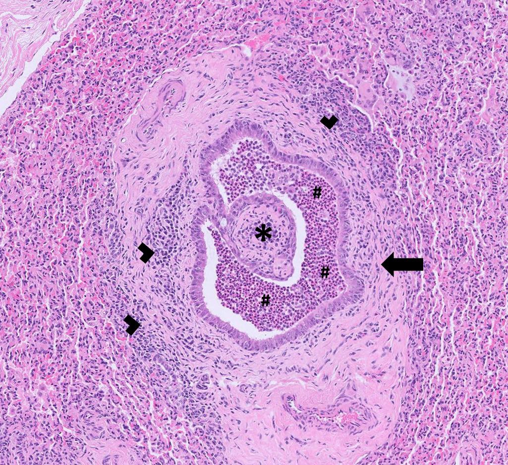
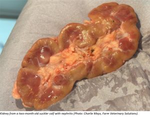
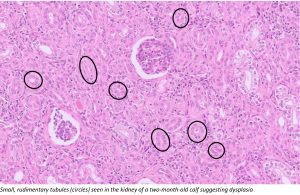
Nephritis was suspected to be the cause of death in a calf that was the second to die suddenly at around two months of age. The calves were noted to be dehydrated prior to death but otherwise there were no premonitory signs. Histopathology identified large areas of significantly compromised renal cortical tissue likely to be associated with renal failure. Although some inflammation was noted, the most striking change observed was the small size and rudimentary nature of the tubules, suggesting a primary dysplasia. Underlying teratogenic (viral or toxic exposure) or perhaps genetic cause(s) were considered. In this case, raised muscle enzymes CK and AST and a low GSH-Px level of 18U/ml RBC was suggestive of possible White Muscle Disease (WMD) in addition to the renal abnormalities. Muscle histopathology is very useful in confirming a diagnosis of WMD; degeneration is usually polyphasic.

Kidney from a two-month-old suckler calfSmall, rudimentary tubules


  • Failure of passive transfer of immunity was identified in a group of calves of which several had died at around a week of age following scouring and respiratory signs. PME had revealed evidence of septicaemia. A recent change in management that involved leaving the calves with the mothers for up to 48 hours was suspected to be the root cause of the problem.
  • Seven adult suckler cows tested positive for antibodies to tick borne fever with high titres indicating recent exposure to Anaplasma phagocytophilum. The reason for testing was not provided in this case.
  • A Proteus sp. was cultured in moderate growth from a urine sample from a dairy cow with cystitis and suspected pyelonephritis that had a poor response to treatment with penicillin/streptomycin antibiotic.

Sheep and goats

Abortion, infertility and mastitis

  • Enzootic abortion was diagnosed as the cause of late abortions and stillbirths on at least 15 occasions this month. Diagnoses were made predominantly on MZN stained smears of placentae and, in some cases, these were confirmed with PCR testing on placentae.
  • Campylobacter fetus was isolated on at least 14 occasions from the stomach contents of lambs aborted up to six weeks early, stillborn lambs and weakly lambs. In at least one case, Campylobacter jejuni was isolated from the stomach contents of an aborted lamb. In another flock, where multiple groups of in lamb ewes were in poor condition and five late term abortions had occurred over an eight day period, Campylobacter coli was isolated from the stomach contents of an aborted lamb; this is a rare but known cause of abortion in sheep.
  • Toxoplasma gondii associated abortion, stillbirths and weakly lambs were confirmed on PCR or foetal fluid serology on at least eight occasions. In one flock, odoriferous off-colour placentae, mummified lambs and very weak lambs were reported.
  • Salmonella 61:k:1,5,7 was isolated on at least three occasions from the stomach contents of aborted or stillborn lambs. In one flock, approximately six full term lambs were born dead and had a foul smell. The ewes were fine up until lambing but become sick after delivering their lambs. In another flock, infected ewes were also scouring.
  • A moderate growth of Streptococcus uberis was isolated in pure culture from the stomach contents of two lambs submitted from one flock. It was not clear if the lambs were aborted by the same ewe but it is likely that this isolate had caused sporadic abortion following ascending infection and opportunistic infection of the gravid uterus.
  • Anaplasma phagocytophilum was detected by PCR in placenta from one of three out of 121 ewes that had aborted in a flock six to eight weeks before the due lambing date. Infection in pregnant ewes (Tick Borne Fever) is a potentially important cause of abortion in sheep. In a second flock with a high barren rate (10%) and low scanning percentage, the presence of ticks was known and the flock had been dipped in November, one month after tupping. Three of the four ewes sampled had a high titre to A. phagocytophilum, suggestive of recent exposure, and one had a low positive titre. In a third flock, two ewes carrying dead lambs had moderate titres to A. phagocytophilum and the other four had low titres.
  • Seroconversion to Border disease virus was reported in at least three barren ewe submissions. In one flock of mules, there was a high barren rate (24/200 empty), with 17/24 gimmers barren. All had been tupped in one group of 200 with four rams and the ewes were in good body condition. Of the seven animals blood sampled, six were seropositive and the seventh had a suspect titre. In another flock, of 42 bought in, in-lamb shearlings, 21 were seropositive for BDV.

Respiratory disease

  • A pregnant ewe died suddenly in the third trimester. The ewe had not been vaccinated against Pasteurella and there was known to be ovine pulmonary adenocarcinoma (OPA) in the flock. On histopathology, the dominant change was severe suppurative bronchopneumonia and severe fibrinosuppurative pleuritis. These inflammatory changes obscured all of the normal lung parenchyma. However, in the background, there were also multifocal areas that most likely reflected concomitant OPA. Animals with OPA commonly present with exercise intolerance and ill thrift. However, if a secondary bacterial pneumonia develops, as here, this can often lead to sudden death. The isolation of Mannheimia haemolytica from lung suggested this was the likely cause of the secondary bacterial bronchopneumonia and pleuritis. Two other cases of OPA were also confirmed on histopathology.

Gastrointestinal disease

  • In February, a total of 124 submissions for Johne’s disease serology and faecal PCR testing were received from sheep and goats, with 30 submissions, including a total of 38 individual samples, testing positive. Cases included a four-year-old Swaledale ewe that was thin, recumbent and had bottle jaw, and a nine-year-old male goat that was thin and still eating and was euthanased as it had been recumbent for 10 days.
  • Cases of parasitic gastroenteritis were confirmed in both sheep and goats, with the presence of high strongyle egg counts. Presenting signs included weight loss, scour and some deaths. In one post weaned goat kid from a herd with a known history of haemonchosis, a strongyle egg count of 10,200 epg suggested this kid was also suffering from Haemonchus infection. A number of cases of treatment failure were also reported on AHWP worming treatment checks.
  • Several cases of fascioliasis were diagnosed on microscopy and coproantigen ELISA, particularly in sheep. Cases included a recumbent, anaemic Black Welsh Mountain ewe that was treated with ivermectin and subsequently died.
  • Type D clostridial enterotoxaemia was suspected following the detection of Clostridium perfringens alpha and epsilon toxin on ELISA testing of small intestinal contents from an adult pygmy goat that died acutely.
  • E. coli antibiotic sensitivity profiles were provided for several suspected cases of watery mouth in neonatal lambs, some of which proved to be multiresistant. The recommended sites for swabbing are the mouth in live lambs and the abomasum in dead lambs.

Skin and eye disease

  • Mycoplasma conjunctivae infection was diagnosed on PCR of ocular swabs on at least three occasions. In all cases, ocular disease appeared to not respond to treatment or recur within days of ceasing treatment.
  • Seroconversion to sheep scab was seen in multiple flocks this month. As in some cases no history was given, it was not clear if positive results were consistent with active infection or persistent antibody levels following successful treatment as positive results can be detected for up to 9 months after successful treatment in some animals.

Systemic and miscellaneous disease

  • Seroconversion to Maedi visna (MV) was reported in several flocks, both on screening and diagnostic submissions. In one flock, where five out of 10 thin ewes screened for MV had been seropositive, a screen of 40 animals revealed that 30 were seropositive, indicating a high rate of infection in the flock.
  • A goat with abscesses was seropositive to CLA. This was the second confirmed case in the herd.
  • Trueperella pyogenes was isolated from a chronic abscess on the face of a three-year-old Texel ewe that was tracking down the animal’s neck.
  • Streptococcus dysgalactiae was cultured from joint swabs from two lambs in a flock in which lambs were going off their legs and were unable to hold up their heads. This is the most common cause of joint ill in young lambs.
  • PME of one of two ill thrifty Jacob lambs found dead revealed liquid gut contents, pulpy kidneys and glucosuria. The lambs were not vaccinated and the detection of Clostridium perfringens epsilon toxin on ELISA testing of intestinal contents was suspicious of pulpy kidney.
  • Listeriosis was diagnosed in a three-year-old Valais Blacknose ewe with histopathology of the brain stem detecting a multifocal suppurative rhombencephalitis.

Brain stem of a sheep with listeriosis


Deficiencies and toxicities

  • Cobalt and vitamin A deficiency were identified in a group of one to seven-year-old Boer goats that had skin scabbing around the eyes, nose and coronary band and had responded poorly to treatment for ectoparasites. Cobalt deficiency was also identified in another Boer goat that had shelly hoof suspected to be secondary to a trace element deficiency.
  • Hypocupraemia was seen in four Dexter cattle of varying ages that were reported to be in poor condition. Copper deficiency was also seen in groups of poor doing Aberdeen Angus calves and suckler cows in poor condition.
  • Manganese levels were very low in all five animals tested in a case of ill thrift.
  • Selenium deficiency was diagnosed in adult suckler cows with long-term weight loss and in ill thriven youngstock with poor growth rates.
  • In one beef suckler herd there were reports of weakly calves at birth and six or seven calves had died. Four of the six cows tested had low copper results and four had marginally low GSH-Px results suggesting possible hyposelenosis.
  • Various combinations of hypocalcaemia/hypomagnesaemia and marginal hypo calcaemia/magnesaemia were seen in cases of down cows both pre and post-calving. In one herd, there had been two sudden deaths in adult cows in the previous two months. Magnesium was tested in five cows from the same group and three of the five were low and one had a marginal result. Four of the five also had low plasma copper. In another herd, samples were submitted from five cows that had all become recumbent within 24 hours of calving. Four of the five were hypocalcaemic and hypophosphataemic, three of the five had magnesium levels less than 0.8mmol/l and so also could be considered marginally low for magnesium.
  • One group of Limousin cows were reported to be having issues with slow calvings and prolapses. Six of six cows tested were marginally hypomagnesaemic. A pooled plasma inorganic iodine result from the group indicated that recent dietary iodine intake was very high, it wasn’t known what, if any, iodine supplementation had been provided.
  • Hypomagnesaemia was identified post mortem on an ocular fluid sample in a case of sudden death in an Aberdeen Angus cow.
  • Blood samples were received from a group of calving heifers that were reported to be in poor condition. Some had calved early, a few calves were reported to be slow and dopey and a couple had died. T4 was very low in the group indicating they were at high risk of clinical disease related to iodine deficiency, copper results were low consistent with copper deficiency and GSH-Px levels suggested marginal selenium status.
  • Low urea levels indicating a deficiency of rumen degradable protein (RDP) were seen on numerous occasions in groups of suckler cows and also in growing animals that presented with poor performance and ill thrift. One group of 40 beef cows were in poor condition and were being fed poor quality silage with no concentrates. All five cows tested had urea levels below the reference range. RDP deficiency can increase the risk of rumenal/ abomasal impaction, especially in cows bearing twins. On another farm, several of a group of bought-in Limousin cross cows with calves at foot showed chronic loss of condition since arrival. They had received anthelmintic and flukicide and been given a trace element bolus. They were reported to have received silage produced from ground with a high lead level. All five cows that were tested had low urea levels and there was evidence of exposure to lead above expected background levels.
  • Evidence of hypocalcaemia and marginal hypomagnesaemia was seen in a group of ewes that had an increased rate of vaginal prolapse pre-lambing.
  • Six ewes from a group of 100 died shortly after moving pastures. They showed signs of drooping ears and depression shortly before death. No lesions were observed at PME of one of the dead ewes. A depressed ewe was blood sampled and was found to be hypocalcaemic.
  • Ewes that had poor nutrition were showing signs of hypoglycaemia and hypocalcaemia on clinical examination. Hypocalcaemia and evidence of ketosis were confirmed on blood samples.
  • A ewe that had to be euthanased after suffering mastitis two weeks post lambing was found to have a high BHB level of 6mmol/l confirming severe ketosis.
  • Vitamin A deficiency was identified in a blind 15-month-old North Devon heifer, a 10-day-old Holstein-Friesian calf with suspected blindness and a one-day-old Holstein-Friesian calf that was born with an undershot jaw and eye abnormalities.

Camelids

Gastrointestinal disease

  • Parasitic gastroenteritis was suspected in a 14-year-old male alpaca that had been losing weight over a few weeks and a strongyle egg count of 1050 epg confirmed the suspicions. A strongyle egg count of 2750 epg was reported in a sick alpaca with a poor condition score and pale mucous membranes, potentially consistent with suspected haemonchosis, although peanut agglutinin testing was not carried out to confirm this. A five-year-old male alpaca with chronic condition loss and loose faeces had a strongyle egg count of 2200 epg. An ill thrifty 12-year-old female alpaca with a suspected tooth root abscess had a strongyle egg count of 2000 epg, 250 epg Nematodirus battus and 600 epg Trichuris (known to be pathogenic in camelids). An approximately seven-month-old male alpaca that was reluctant to move and had a slightly raised temperature and pale conjunctivae had a strongyle egg count of 400 epg, 500 epg Trichuris and a coccidial oocyst count of 36,200 opg, consistent with concurrent coccidiosis.
  • A faecal oocyst count of 30,650 opg Eimeria macusaniensis was reported in a recumbent, elderly alpaca in poor body condition and with pale mucous membranes, despite a good appetite. This large coccidian is the only Eimeria species that tends to cause clinical coccidiosis in all ages of alpaca. A recumbent adult alpaca was also found to have an E. macusaniensis count of 2000 epg and a strongyle egg count of 1700 epg, consistent with concurrent parasitic gastroenteritis.
  • Fascioliasis was diagnosed on faecal microscopy in a 12-year-old stud male alpaca with severe weight loss and in a five-year-old male alpaca with an unknown clinical history.
  • Giardiaisis was diagnosed as the likely cause of persistent scour in a Bactrian camel. Campylobacter jejuni was also isolated from faeces but was of unclear significance as this is often found in the faeces of healthy animals.

Skin and eye disease

  • A herd of 50 alpacas were affected with widespread pruritus, skin crusting and alopecia. The ventrum and distal limbs were worst affected and several animals had died or been euthanased over the previous few months. Skin microscopy taken at the PME of one affected animal detected numerous adults, nymphs and larvae of Psoroptes mites. Mites were also seen on histopathology of skin samples and there was evidence of secondary pyoderma. Although of the same genus as sheep scab mites, those found in camelids are not believed at present to be a reservoir of infection for sheep.

Mites in the skin

Other Species

  • F4 (K88) associated E. coli was detected in the faeces of seven-day-old piglet in a litter of piglets that started to scour at less than four days of age and eight out of the 16 in the litter had died. The E. coli was multi-resistant. F4 antigen was also detected in a faeces sample from a litter of three six-week-old pigs that were presumed to be scouring.
  • Fascioliasis was diagnosed in three adult reindeer in a herd using coproantigen ELISA.

Follow us on Facebook

https://www.facebook.com/Axiomfarmvetlab/

This newsletter is sent by e-mail to each vet practice but if you would like a copy sent to your individual e-mail account please contact us at dsfarm@axiomvetlab.co.uk and we can add you to our circulation list.

Related Posts