Email Us
admin@axiomvetlab.co.uk

admin@axiomvetlab.co.uk
(01626) 355655

This newsletter is sent by e-mail to each vet practice but if you would like a copy sent to your individual e-mail account please contact us at dsfarm@axiomvetlab.co.uk and we can add you to our circulation list.
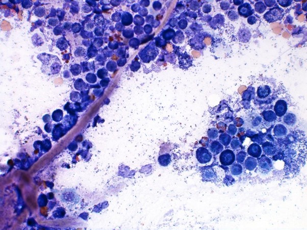
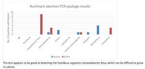
We have results for the first fifty-two submissions from our new ruminant abortion PCR package, which can detect ten different causes. An agent has been detected in 65% of the small ruminant abortion cases (15/23 submissions, one submission had two agents present). For bovine submissions an agent has been detected in 31% of the 29 submissions tested to date. Both of these are an improvement on the usual diagnostic rates for abortions despite the fact that a number of abortions this year are likely to be due to Schmallenberg virus (which the package does not cover) and unfortunately in some areas bluetongue is also having a significant impact through causing abortions, infertility and dummy calves.
If there are concerns about Schmallenberg virus then submit foetal fluid for serology (usually test this first as significantly cheaper to do so) but if negative consider then testing for the virus, which requires a piece of umbilicus/placenta/brain/brainstem/spleen. Cases can be virus positive or antibody positive or sometimes both.
For the abortion PCR package (test code FABORT) submit:
Foetal stomach contents, liver & brain or
vaginal swab and ideally also a piece of cotyledon (without faecal contamination).
We have detected evidence of low RDP intakes in a number of herds this winter where the history has been of youngstock or adult cattle that are failing to thrive. It is also a relatively common cause of ill thrift in bulls so it appears that some bulls are being underfed protein, particularly if they are still growing. This year it seems that some silages are lower in protein than had been thought to be the case. If RDP intakes are suboptimal after calving then milk yields and fertility can also be adversely affected. Low RDP intakes are detected through testing blood samples for urea, which can be done at a low cost of five samples for £24 (i.e. £4.80 per sample for five or more). We are not just looking for low blood urea results but suboptimal levels. However, if blood urea levels are low in late pregnancy then colostral quality (and potentially quantity) are likely to be adversely affected.
We have two testing options for winter dysentery – paired coronavirus serology to check for rising antibody levels or PCR testing for the presence of virus using faecal samples. The vast majority of adult cattle have detectable antibodies (though immunity does not appear to be complete) therefore in order to demonstrate recent exposure to coronavirus it requires paired serology to check for rising antibody levels. Blood samples should be collected 2-4 weeks apart. Alternatively, for a quicker result, faeces can be tested for the virus using a PCR test. Virus tends to only be shed for a very short time following infection so it is important that new cases are sampled. Although the PCR test has not been validated for testing of pooled samples we seem to have had better success with detecting it in pooled rather than individual faecal samples. With it being a PCR test it is a sensitive test and by sampling multiple animals it increases the chance of detecting the virus. We would recommend no more than three faecal samples in a pool to reduce the risk of over dilution with negative samples, which could compromise sensitivity.
Please note that we are now participating in the worm egg counting part of the AHWP for sheep. However, we are unable to post out sampling kits. Consumables can be ordered from us in the usual way. WHEN SUBMITTING POST TREATMENT SAMPLES, PLEASE ENTER THE ACCESS (REPORT) NUMBER FOR THE PRE-TREATMENT SAMPLE RESULTS AS A PREVIOUS REFERENCE ON THE SUBMISSION FORM. We can then provide you with a % change in the strongyle egg count after treatment.
We are a UKAS accredited lab and provide ISO17025 accredited tests so we can carry out any of the follow up endemic disease testing for both cattle and sheep. The diseases and conditions to be sampled for sheep include: Border disease (BD), caseous lymphadenitis (CLA), enzootic abortion of ewes (EAE), Johne’s disease, Maedi Visna (MV), toxoplasmosis, tick-borne fever, pulpy kidney, lamb dysentery, ewe nutrition status, lamb nutrition status & trace elements.
The above list is only an example of scenarios, however where we experience additional time and cost in processing poorly submitted samples we reserve the right to charge an additional fee towards the admin time incurred e.g. for blood samples that fee would be 25p per tube.
Sending us a complete and accurate ID reference list with your herd/multiple animal submissions for checking and booking in your samples is an essential part of the procedure.
This is facilitated by using the detachable bar codes supplied on our preferred serum gel tubes. Simply record the animal ID on the Axiom submission form (or your own list/table) then remove the barcode strip from the tube and place it alongside the corresponding ID in order for us to associate each specific tube with the relevant animal. There is no need to write out the bar code numbers by hand on the list.
Please remember always to provide such a list and, when using a Field Kit for larger herd submissions, be sure to populate the polystyrene rack IN THE SAME ORDER AS YOUR ID LIST. This means that we will be able to check, process, and forward your samples to the relevant laboratory department for testing in a far more efficient manner, in turn allowing us to deliver your results in the shortest possible timeframe.
Please provide your name, practice, the farmer and farm names so that we can link the photos to the relevant submission and please also indicate which Axiom vet you discussed the case with. We may wish to use some of the photos in our newsletter so please indicate if you are not happy for this to be done. All cases are anonymised and credited to the submitting vet. Please note that this number is just for sharing photos. If you wish to discuss a case for which you do not have photos, please ring 01626 357776 as usual.
We are an accredited lab for the Welsh BVD eradication programme. BVD antibody and antigen results will be uploaded if samples are submitted on a BVD Cymru form. As was the case with BVD Free England, there is a small charge for the uploading of the results of 50p per sample for BVD antibody testing and 25p for a BVD antigen test.
Please note that all fields on the BVD Cymru submission form must be completed (including the keeper’s phone number and email address) otherwise there is a block on the results uploading.
Our Johne’s & Neospora Monitoring programmes give farmers access to discounted test rates for whole herd or regular batch testing. There is no membership fee and no set rules to follow. Johne’s serology is from £4.25 per sample and Neospora serology costs from £5.90 per sample. It works out cheaper than testing through a CHECS cattle health scheme so is ideal for herds that are testing for disease control and management purposes. Advice is provided in the lab report and farmers can be e-mailed a copy if required. Our turnaround times are very fast– often same day but within three working days for both tests. Batch testing herds also get their results in a cumulative spreadsheet. Contact us for more information at dsfarm@axiomvetlab.co.uk or on 01626 357776.
In order to avoid any unnecessary confusion, please can we ask that submission forms are only sent in with the samples and not in advance of the samples. Thank you for your cooperation.
You may find this is a more efficient way of making requests than phoning the farm team, saving you time in your busy day. Our farm team also find it a more efficient way of dealing with your requests.
The email address for test requests is: DSFarm@axiomvetlab.co.uk
Cattle
Abortions
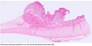
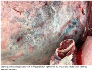
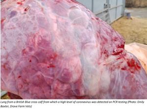
Respiratory disease
Gastrointestinal disease
Systemic and miscellaneous disease
Abortion, infertility and mastitis
Respiratory disease
Gastrointestinal disease

Skin and eye disease
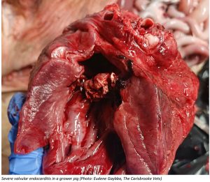
Systemic and miscellaneous disease
Deficiencies and toxicities
Gastrointestinal disease
Systemic and miscellaneous disease

Farm May Newsletter 2026 Dedicated farm line – 01626 357776 Follow us on Facebook https://www.facebook.com/Axiomfarmvetlab/…
Farm April Newsletter 2026 Dedicated farm line – 01626 357776 Follow us on Facebook https://www.facebook.com/Axiomfarmvetlab/…
Farm March Newsletter 2026 Dedicated farm line – 01626 357776 Follow us on Facebook https://www.facebook.com/Axiomfarmvetlab/…
Download a PDF
The great explorer of the truth, the master-builder of human happiness no one rejects dislikes avoids pleasure itself because it is pleasure but because know who do not those how to pursue pleasures rationally encounter consequences that are extremely painful desires to obtain.
Read More