Farm November Newsletter 2024

Dedicated farm line – 01626 357776

This newsletter is sent by e-mail to each vet practice but if you would like a copy sent to your individual e-mail account please contact us at dsfarm@axiomvetlab.co.uk and we can add you to our circulation list.


Animal Health & Welfare Pathway – worm egg counts now available

Please note that we are now participating in the worm egg counting part of the AHWP for sheep. However, we are unable to post out sampling kits. Consumables can be ordered from us in the usual way. WHEN SUBMITTING POST TREATMENT SAMPLES, PLEASE ENTER THE ACCESS (REPORT) NUMBER FOR THE PRE-TREATMENT SAMPLE RESULTS AS A PREVIOUS REFERENCE ON THE SUBMISSION FORM. We can then provide you with a % change in the strongyle egg count after treatment.
We are a UKAS accredited lab and provide ISO17025 accredited tests so we can carry out any of the follow up endemic disease testing for both cattle and sheep.

The diseases and conditions to be sampled for sheep include:
Border disease (BD), caseous lymphadenitis (CLA), enzootic abortion of ewes (EAE), Johne’s disease, Maedi Visna (MV), toxoplasmosis, tick-borne fever, pulpy kidney, lamb dysentery, ewe nutrition status, lamb nutrition status & trace elements.

Incorrectly submitted samples

Samples received into the lab that are incorrectly packaged require more time and equipment to process and in some instances present an unnecessary Health and Safety risk to our Lab staff. Examples of incorrectly packaged samples include:

  • Samples submitted in rubber gloves rather than the correct sample pots, e.g. faecal samples.
  • Non serum gel tubes for clotted blood samples (we would ask you to use serum gel tubes provided by Axiom Laboratories which substantially decrease sample processing time and reduce the need for additional plastic consumables as the analyser can test direct from these tubes).
  • Cases that contain multiple samples from different animals that are either unlabelled or that have been packaged out of the numerical sequence written on the accompanying submission form.
  • No list of animal IDs and tube numbers received with herd or flock screens. This list is essential for booking in samples in the office.
  • Where needles have been included in the packaging along with the samples.
  • The submission of whole animals and foetuses as we do not have a PM room to handle these.

Continuing to submit incorrectly packaged samples affects our ability to get results to you quickly and efficiently while keeping our prices as low as possible.

The above list is only an example of scenarios, however where we experience additional time and cost in processing poorly submitted samples we reserve the right to charge an additional fee towards the admin time incurred e.g. for blood samples that fee would be 25p per tube.

Would you like to send the Axiom farm vets photos of PMEs to allow us to assist with sample selection and diagnosis? Please send photos by WhatsApp to 07944 649222.

Please provide your name, practice, the farmer and farm names so that we can link the photos to the relevant submission and please also indicate which Axiom vet you discussed the case with. We may wish to use some of the photos in our newsletter so please indicate if you are not happy for this to be done. All cases are anonymised and credited to the submitting vet. Please note that this number is just for sharing photos. If you wish to discuss a case for which you do not have photos, please ring 01626 357776 as usual.

BVD Cymru accredited lab

We are an accredited lab for the Welsh BVD eradication programme. BVD antibody and antigen results will be uploaded if samples are submitted on a BVD Cymru form. As was the case with BVD Free England there is a small charge for the uploading of the results of 50p per sample for BVD antibody testing and 25p for a BVD antigen test.

New multiplex PCR test for investigating ruminant abortions.

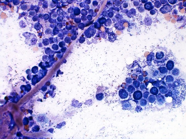
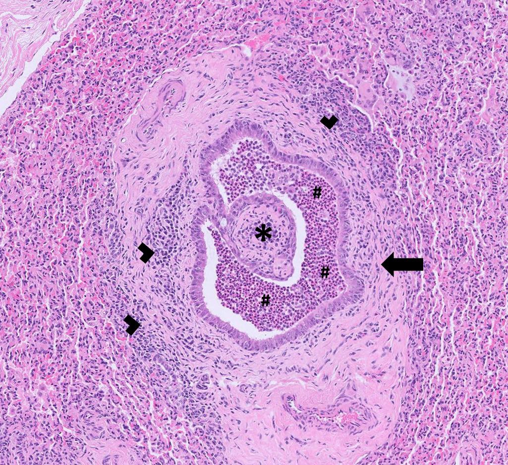
The diagnostic rate for bovine abortions through veterinary laboratories has always been relatively low. There can be a number of reasons for this – the cost of testing for the large range of possible causes makes it cost prohibitive to do a comprehensive screen, the degree of autolysis, lack of availability/testing of placenta and of course some will have been aborted for non-infectious reasons e.g. nutritional, physical or genetic reasons. With sheep abortions the diagnostic rate is higher but screening is usually limited to the most common causes. In an attempt to increase the diagnostic rate of infectious causes of ruminant abortions we are introducing a ten agent multiplex PCR test. It will screen for the following agents: Anaplasma phagocytophilum (TBF), Campylobacter fetus, Chlamydophila spp. (includes EAE), Coxiella burnetii (Q fever), Leptospira spp., Listeria monocytogenes, Neospora caninum, Salmonella spp., Toxoplasma gondii & Brucella sp. These agents are often found systemically in a foetus but can vary as to which viscera they are found in and also the levels at each site. Placenta is often a very good sample to include in a pool of tissues however if it is contaminated with faeces this can be detrimental. Faeces can contain Campylobacter fetus and also Chlamydia pecorum (a Chlamydophila sp. that does not cause abortions) which could lead to a mis-diagnosis. If placenta is faecally contaminated then it is better to sample tissues, using clean instruments and gloves, from within the carcase. The following fresh tissues, which we will test as a pooled sample, are likely to give a very good chance of detecting the above agents if they are present:
Brain (or uncontaminated placenta) & liver & foetal stomach contents (FSC)
Or if no foetus available:
Vaginal swab (plain) & uncontaminated placenta
Clean the vulva with paper towel and part the vulval lips to avoid faecal contamination of the swab and take the sample from the ventral aspect of the cranial vagina. Please note that if only a vaginal swab is submitted (taken up to five days after parturition) and no placenta is sampled then testing is unlikely to detect the presence of Neospora, Toxoplasma and probably also TBF – though serology could be done for these agents instead.
The screen does not include BVD virus (foetal fluid can be tested for BVD antigen and antibodies or there is the more expensive PCR test using fresh spleen or liver) or IBR (we have a PCR test using fresh liver, as an add on).
Please note that the screen also includes Brucella species so if a positive result is obtained for this APHA would need to be informed, with it being a notifiable disease.

Discounted test rates for Johne’s disease & Neospora herd screens or regular batch testing

Our Johne’s & Neospora Monitoring programmes give farmers access to discounted test rates for whole herd or regular batch testing. There is no membership fee and no set rules to follow. Johne’s serology is from £4.25 per sample and Neospora serology costs from £5.90 per sample. It works out cheaper than testing through a CHECS
cattle health scheme so is ideal for herds that are testing for disease control and management purposes. Advice is provided in the lab report and farmers can be e-mailed a copy if required. Our turnaround times are very fast– often same day but within three working days for both tests. Batch testing herds also get their results in a cumulative spreadsheet. Contact us for more information at dsfarm@axiomvetlab.co.uk or on 01626 357776.
In order to avoid any unnecessary confusion, please can we ask that submission forms are only sent in with the samples and not in advance of the samples. Thank you for your cooperation.

Did you know that you can email us any additional test requests for your submissions?

You may find this is a more efficient way of making requests than phoning the farm team, saving you time in your busy day. Our farm team also find it a more efficient way of dealing with your requests.
The email address for test requests is: DSFarm@axiomvetlab.co.uk

Discounted test rates for Johne’s disease & Neospora herd screens or regular batch testing.

Our Johne’s & Neospora Monitoring programmes give farmers access to discounted test rates for whole herd or regular batch testing. There is no membership fee and no set rules to follow. Johne’s serology is from £4.25 per sample and Neospora serology costs from £5.90 per sample. It works out cheaper than testing through a CHECS cattle health scheme so is ideal for herds that are testing for disease control and management purposes. Advice is provided in the lab report and farmers can be e-mailed a copy if required. Our turnaround times are very fast– often same day but within three working days for both tests. Batch testing herds also get their results in a cumulative spreadsheet. Contact us for more information at dsfarm@axiomvetlab.co.uk or on 01626 357776.
In order to avoid any unnecessary confusion, please can we ask that submission forms are only sent in with the samples and not in advance of the samples. Thank you for your cooperation.

Did you know that you can email us any additional test requests for your submissions?

You may find this is a more efficient way of making requests than phoning the farm team, saving you time in your busy day. Our farm team also find it a more efficient way of dealing with your requests.
The email address for test requests is: dsfarm@axiomvetlab.co.uk

Would you like to send the Axiom farm vets photos of PMEs to allow us to assist with sample selection and diagnosis? Please send photos by WhatsApp to 07944 649222.

Please provide your name, practice, the farmer and farm names so that we can link the photos to the relevant submission and please also indicate which Axiom vet you discussed the case with. We may wish to use some of the photos in our newsletter so please indicate if you are not happy for this to be done. All cases are anonymised and credited to the submitting vet.

Interesting cases in October


Cattle


Abortions, cryptorchidism

  • Several cases of Neospora infection were detected by PCR in aborted fetuses. To confirm neosporosis as the cause of abortion, histopathology of foetal heart and ideally foetal brain is required.
  • Trueperella pyogenes was isolated from stomach contents of aborted foetuses in two dairy herds. In one herd, a six-year-old Holstein cross cow aborted at six month’s gestation. In the second herd, the second foetus aborted in 72 hours was sampled and this also had a positive BVD antigen ELISA result, potentially consistent with in utero BVD virus infection (e.g. following a transient BVD viraemia) or the use of a live BVD vaccine in a pregnant animal, although a false positive result also could not be ruled out. If there was transient BVD viraemia, this could have caused immunosuppression and increased the risk of haematogenous spread of T. pyogenes from a septic focus, e.g. lung, liver or foot abscess. It was advised that further investigation of the herd’s BVD status was carried out, including bulk milk serology and virus PCR testing, and antibody check tests on youngstock groups.
  • Listeria monocytogenes was isolated from the stomach contents of an Aberdeen Angus foetus aborted at three months’ gestation. Typically, abortion is associated with the ingestion of spoiled or soil contaminated silage, or grazing on muddy ground.

Respiratory disease

  • IBR was confirmed by PCR on at least seven occasions in October, with cases seen in five-month-old calves to adult cows. In one case, a group of heifers were exhibiting respiratory and ocular signs, including purulent ocular discharge, and also lesions consistent with vulvovaginitis. IBR was detected by PCR from both pools of ocular swabs and both pools of vaginal swabs tested.
  • PI3 was detected by PCR on at least seven occasions, the majority of cases being confirmed on pooled nasopharyngeal swabs. However, in one case, one of a group of six-month-old calves that had arrived on farm one month earlier and had been showing signs of pneumonia for one to two weeks, declined rapidly and died, despite treatment. On histopathology, there was evidence of a severe necrotising bronchiolitis consistent with a previous pneumotropic viral infection such PI3. This had been followed by a secondary bacterial infection which at this stage predominated and likely accounted for the rapid decline. In a second case, RSV was also detected with PI3 in one of two pools of nasopharyngeal swabs submitted from two-week-old calves.
  • RSV was detected by PCR on at least a further eight occasions. In one case, a six-month-old Charolais cross calf in a batch of coughing, pneumonic animals died quickly over 24 hours. Lungworm was suspected but were not seen on PME, which revealed gross pathology particularly in the cranioventral lobes. Histopathology detected extensive necrosuppurative bronchointerstitial pneumonia with interlobular emphysema consistent with RSV infection. There was also suggestion of a secondary bacterial infection. In one herd, RSV infection was confirmed on paired serology in one of five dairy cows with presumed respiratory disease.
  • At least four cases of a bacterial bronchopneumonia with underling lesions likely to be consistent with initial pneumotropic viral infection (bronchiolitis fibrosa obliterans) were diagnosed on histopathology. In one case, a five-month-old Holstein-Friesian calf that had extensive consolidation of the ventral lungs, no viruses could be detected on PCR but the chronic nature of the lesions was the likely explanation for this. Histophilus somni, Mannheimia haemolytica and Pasteurella multocida were detected by PCR and Bibersteinia trehalosi was isolated on culture; any or all of these may have been contributing to the secondary bacterial infection. Mycoplasma bovis was also detected by PCR and, although changes typical of a primary virulent Mycoplasma bovis infection (eosinophilic coagula occluding airways) were not seen, there was minimal to mild plasmalymphocytic inflammation which would support a concurrent Mycoplasma species involvement.
A chronic airway from a calf with bovine respiratory disease. Arrow = peri-bronchiolar fibrosis and * = bronchiolitis fibrosis obliterans which suggest previous necrotising damage to the airways, most often in young calves caused by a virus (e.g. BHV-1, RSV or PI3). Arrow heads = lymphocytes and plasma cells (peri-bronchiolar cuffing) which can suggest concurrent Mycoplasma spp infection or chronic antigenic stimulation. # = neutrophils in the airway suggesting a secondary bacterial bronchopneumonia
  • A 30-hour-old Highland calf, whose birth was not observed, was weak and could only stand for short periods with assistance. On examination, the calf was breathing heavily, with an increased respiratory rate, and died following the examination. On PME, the lungs were diffusely dark and histopathology was consistent with broncho-interstitial type pneumonia following in utero exposure to an infectious agent/antigenic stimulation. Prominent lymphoid follicular hyperplasia around bronchioles (‘cuffing pneumonia’) suggested either Mycoplasma or Ureaplasma involvement, and, as the inflammatory process was also somewhat neutrophilic, this raised the possibility of involvement of other bacteria (for example secondary to a placentitis).
Prominent lymphoid follicles (arrows) in the lungs of a neonatal Highland calf with respiratory signs
  • Post mortem tissues were received from a 13-month-old dairy heifer that died acutely with no premonitory clinical signs. Changes in the lung were consistent with a necrotising bacterial pneumonia and any or all of the bacteria detected by PCR in fresh lung (Histophilus somni, Mannheimia haemolytica, Pasteurella multocida and Mycoplasma bovis) may have been implicated. In addition, there was evidence of myocarditis and epicarditis most likely due to Histophilus somni. The mononuclear population of inflammatory cells seen in the heart suggested that, although death appeared sudden, this may have been a slightly longer standing condition.
  • Mycotic pneumonia was confirmed on histopathology of lung tissue from a freshly calved heifer (no further history provided), with the finding of widespread acute and severe necrotising pneumonia associated with abundant fungal hyphae. The distribution of the fungal hyphae was most suggestive of a haematogenous spread following inhalation of fungal spores from heavily contaminated food, although infection acquired from the alimentary tract following ingestion of mouldy food was also a possibility. In the majority of cases, Aspergillus species are implicated however other organisms including Mucor, Rhizopus, Absidia and other zygomycetes should also be considered.
  • Several cases of lungworm were confirmed on Baermann’s testing of faeces samples from animals as young as five-months-old, clinical signs including coughing, increased respiratory noise and ill thrift. Several animals with respiratory signs were also seropositive for lungworm.
  • Salmonella Dublin was isolated from lung swabs from pneumonic calves in at least three herds. In one case, in Holstein-Friesian calves less than two months of age, the calves were also scouring.

Gastrointestinal disease

  •  Coccidiosiswas confirmed in a six-month-old pyrexic Hereford calf that had passed bloody scour for a few days. A faeces sample had a coccidial oocyst count of greater than 50,000 opg of which 60% were of a pathogenic Eimeria species, predominantly E. zuernii. A second case was confirmed in an ill thrifty calf with loose faeces that had a count of 5900 opg of which three quarters were pathogenic Eimeria species, predominantly E. alabamensis. A third case was seen in a shed of ill thrifty dairy and dairy beef cross calves with the finding of a count of 6200 opg in one pen, of which over half were identified as pathogenic Eimeria species (a mix of Eimeria zuernii, E. bovis and E. alabamensis). Other pens had lower counts but, as clinical signs can precede patency, these may still have been of significance. A fourth case was seen in a group of 12-week-old Holstein-Friesian calves that were failing to put on weight; coccidial oocyst counts of individual samples were between 100 and 550 epg and a pooled sample had 100% Eimeria bovis on speciation.
  • High pepsinogen levels associated with significant abomasal parasitism were reported in ill thrifty animals also often presenting with scour. High strongyle egg counts consistent with parasitic gastroenteritis were also reported in several animals. In one case, a thin seven-month-old dairy heifer with loose faeces was brought inside and declined, developing watery diarrhea before dying. 1200 epg strongyles and 200 epg Nematodirus sp. were found in a faeces sample from the animal.
  • Cases of fasciolosis were diagnosed on both fluke microscopy and coproantigen ELISA, including a wasting Charolais bull that was also confirmed as having Johne’s disease on faecal PCR testing and in some coughing spring born dairy calves.
  • In October, a total of 524 submissions for Johne’s disease serology and faecal PCR testing from cattle were received, with 180 submissions, including a total of 415 individual samples, testing positive. Cases were seen in animals as young as 18 months of age and also in at least two beef bulls.
  • Salmonella Dublin was isolated from at least nine faeces samples from scouring animals, ages ranging from neonatal calves to adult cattle. In one Friesian cow, that had been calved for a week and had milk drop and scour, in addition to S. Dublin being isolated, there was a low positive result on PCR for MAP. This could be due to the animal having Johne’s disease, transient passage of MAP, temporary shedding in a recently challenged animal or a false positive result. Salmonella Typhimurium was isolated from faeces samples from at least three scouring animals including a Jersey cow two weeks off calving that developed watery scour and pyrexia (T 39.2oC). Salmonella Mbandaka was isolated from an adult Holstein with presumed scour, Salmonella Montevideo was isolated from faeces from scouring Holstein-Friesian cows and Salmonella Newport was isolated from faeces from recently calved, scouring Holstein-Friesian cows with milk drop. A further case of Salmonella Indiana infection was confirmed in a pre-weaned beef calf in a herd in which a number of cases of this rare serotype in cattle had been confirmed in recent weeks.
  • Post-mortem tissues were received from a 10-day-old dairy calf. Calves on the unit were fed on whey milk powder after receiving two colostrum feeds. However, they were not sucking well and subsequently dying. This calf was euthanased and, on PME, there was evidence of milk in the rumen, a distended abomasum with no milk clots and empty intestines. Histopathology revealed an acute necrohaemorrhagic and fibrinosuppurative abomasitis with abundant gram positive rods consistent with clostridial abomasitis. A second case was confirmed on histopathology in a Holstein cross calf in another herd and here there was also evidence of peritonitis, presumably secondary to perforation of the abomasal wall. A number of predisposing factors are thought to be involved in abomasitis in young calves including compromised immunity from failure of passive transfer, bacterial infection of the abomasal wall, and vitamin and mineral deficiencies.
Lung
Clostridial abomasitis in a 10-day-old dairy calf (Photos: Reuben Newsome, Fosse Way Farm Vets)

Skin and eye disease

  • Dermatophilosis was confirmed on culture of a hair pluck and scab taken from one of a group of weaned Holstein-Friesian calves with scabby skin, ears, withers and shoulders. Part of the group had been born in very wet weather and hair and scab had pulled up very easily, raising suspicions of dermatophilosis.
  • Pediculosis was diagnosed in one animal with the identification of the biting louse Bovicola bovis on skin microscopy.
  • Trueprella pyogenes was isolated from an abscess over the elbow of one animal that was refractory to treatment – this bacterium is known to cause pyogenic infections.

Systemic and miscellaneous disease

  • A total of 235 submissions were received for BVD antigen testing in cattle in September, with 16 submissions, including a total of 41 individual samples, testing positive. Cases of Mucosal Disease included a seven-month-old poor doing Devon x Hereford heifer calf with scour and pneumonia and a Holstein cow with tenesmus, severe scour and odoriferous, haemorrhagic faeces.
  • MCF was confirmed on PCR testing in a six-month-old calf that had pyrexia, acute onset corneal oedema, dermatitis and mucosal ulceration.
  • Babesiosis was confirmed on a haemoparasite screen and on PCR of an EDTA sample from a seven-year-old Hereford cow in Suffolk. Three cows had died since moving fields the previous week which had presumably brought them into contact with ticks for the first time. This cow became recumbent but did respond to treatment with calcium, magnesium and phosphorus. Three further cases were confirmed in Somerset. The first was a beef cow that had been treated for redwater but showed no improvement over the next three days. The other two cases were in the same suckler herd- a recumbent cow and a lethargic bull that was intermittently recumbent. Both animals had very low temperatures, firm faeces and the bull also had a marked azotaemia (urea 66 mmol/l, creatinine 1667 μmol/l).
  • A dairy cow in its fifth lactation presented with milk drop, scour and mild bottle jaw. The cow was hypoalbuminemic but testing for Johne’s and Salmonella was negative and the animal was unresponsive to treatment. It was found to have an enlarged smooth left kidney on rectal examination and, as its condition deteriorated, it was euthanased. Histopathology of the kidney detected amorphous, extracellular, hyaline to finely fibrillar material obscuring glomeruli, seen within the medullary interstitium and additional rarely within walls of blood vessels. This showed salmon pink staining with Congo Red special stain, consistent with renal amyloidosis. In cattle this is almost always secondary to chronic inflammation and can be associated with mastitis, metritis, arthritis, pododermatitis, udder cleft dermatitis and other conditions.
An unrelated case of bovine renal amyloidosis
  • A 17 –day-old Limousin cross calf had a two-day history of dullness, tachypnoea and loss of interest in suckling, with pyrexia (39.7 to 40oC) that was poorly responsive to meloxicam. On the second day, the calf was bleeding from the sites of injections given the day before, there were small pinpoint wounds on eyelids and bleeding from other sites over the body with no obvious signs of trauma. The calf was euthanased due to welfare concerns. Histopathology of bone marrow detected trilineage (erythroid, myeloid and megakaryocytic) hypoplasia. This presentation is most commonly seen in immune-mediated pancytopenia (bovine neonatal pancytopenia), which has been attributed to the use of a particular BVDV vaccine in the past. However, investigations at that time, and since, have identified a small number of animals presenting with identical clinical signs and histology and no associated vaccination, suggesting other aetiological causes for the immune-mediated disease process. Other potential causes of bone marrow suppression include BVD viraemia, nutritional factors and medications. BVD was ruled out by PCR on a pre-mortem blood sample.

Sheep and goats

Respiratory disease

  • Lung lesions typical of Mycoplasma ovipneumoniae infection (atypical pneumonia) were identified by histopathology in tissue from a lamb which was one of five seven-month-old lambs to die in the previous week. Multifocal extensive chronic bronchointerstitial pneumonia with bronchiolar epithelial hyperplasia, cuffing and bronchiolitis fibrosa obliterans was observed. Mycoplasma ovipneumoniae infection is typically spread amongst
    young lambs in housed environments such as the lambing shed and will cause depressed growth rates and increased susceptibility to other pulmonary infections, which are often the cause of apparent sudden deaths.
  • Mannheimia haemolytica was cultured from lung tissue in a case where there had been approximately thirty deaths in a group of five hundred sheep. Significant pulmonary consolidation was observed at post-mortem.
  • Bibersteinia trehalosi was cultured from lung tissue from a lamb which was one of three per-acute deaths to occur three weeks after the animals were bought-in and placed on good pasture. Lung consolidation and haemorrhages in the lungs and visceral organs were noted at post mortem examination.
  • Histophilus somni was isolated from lung tissue from a ram. Although more commonly identified in clinical samples from cattle, H. somni is also known to affect small ruminants.
  • Fibrinosuppurative pneumonia was the cause of death identified by histopathology of lung from a pygmy nanny goat. Pneumonia and pleurisy were observed grossly over the cranial lung lobes. Four goats had recently died in the herd.
  • Fusobacterium necrophorum and Streptococcus ovis were isolated from abscess material from a ram lamb in good condition which died suddenly and was found to have evidence of severe pleurisy and abscesses in the lungs, liver and spleen at post mortem. Fusobacterium necrophorum is recognized as the cause of hepatic necrobacillosis in sheep and Streptococcus ovis is also associated with infections in sheep and therefore both isolates were considered likely to be clinically significant in this case.
  • Fusobacterium necrophorum and Helcococcus ovis were isolated from one of multiple abscesses seen in the lungs of a thin cull ewe. Helcococcus ovis has been found previously in lung lesions in sheep and was considered to be of possible significance in this case.
  • Severe suppurative pneumonia was identified by histopathology in a sheep which was found to have a pyothorax at postmortem.

Gastrointestinal disease

  • In October, a total of 139 submissions for Johne’s disease serology and faecal PCR testing from sheep and goats were received, with 20 submissions, including a total of 30 individual samples, testing positive.
  • Haemonchosis was confirmed (using the peanut agglutinin test) or suspected on more than 20 submissions this month with evidence of anthelmintic treatment failure also seen on a number of occasions.
  • Fascioliasis was diagnosed by microscopy or coproantigen ELISA in more than 20 cases, with eggs or antigen detected in samples from both animals showing no apparent clinical signs and also affected animals mainly with condition loss or submandibular oedema. Pooled liver fluke serology was positive on several occasions including strongly positive in first season grazing ewe lambs.
  • Salmonella 61:k:1,5,7 was isolated in cultures from samples from a farm where 30 ewe deaths had occurred in just over two weeks. Black disease (Infectious Necrotic Hepatitis due to Clostridium novyi) had been previously diagnosed on the same farm. Salmonella 61:k:1,5,7 may be found in both healthy sheep and in sheep with clinical salmonellosis. This is the most commonly isolated Salmonella serotype in sheep. In cases of sudden death or diarrhoea, it is often identified in combination with other pathogens.

Skin and eye disease

  • Bibersteinia trehalosi was isolated from a fluid sample aspirated from the side of the neck in a six-month-old Wiltshire horn sheep. Cytology of the fluid revealed evidence of neutrophilic inflammation and bacterial infection. B. trehalosi is more commonly associated with respiratory and systemic infections in sheep but was considered to be of possible clinical significance in this case.
  • A Staphylococcus aureus isolate, which showed multiple resistance to antimicrobials on sensitivity testing, was isolated from a swab from the ear pinna of a ewe presenting with dermatitis.
  • Eosinophilic dermatitis was determined to be the cause of pruritic, scabby lesions on the pastern areas of a two-year-old Dorset cross Dutch spotted ewe. No improvements were noted after treatments for ectoparasites and bacterial skin infections. Histopathology identified a hyperplastic and eosinophilic dermatitis. A hypersensitivity to fly bites was considered a possibility given the eosinophilic component. On a second farm skin crusting and itchiness was observed around the ear pinnae and peri-orbitally in an adult Norfolk ewe and eosinophilic dermatitis was observed on histopathology. It was noted that only the dark faced sheep in the flock were affected which often seems to be the case with fly bite hypersensitivity.

Systemic and miscellaneous disease

  • Border disease viraemia was detected in one of two bought-in shearlings by PCR testing. A high Ct value of 41 indicating a low level of virus was detected and therefore this was possibly a case of transient infection. Border disease virus was also detected in a pooled sample from aged easycare ewes of which one viraemic animal was detected on individual testing using the BVD antigen ELISA.
  • Histopathology samples were submitted from a ewe lamb which had presented with persistent pyrexia which failed to respond to treatment. At PME, white lesions were observed on the liver and the dorsal margin of the liver had a mottled appearance. The kidneys were pale yellow in appearance and also had white lesions in the cortex. Histopathology identified a granulomatous pneumonia, hepatic infarcts and evidence of vasculitis. The unusual appearance raised the suspicion of possible OvHV-2 infection and the samples were submitted for ISH (in-situ hybridisation) testing which identified strong intra-nuclear labelling of lymphocytes in the perivascular cuffs (positive for OvHV-2). Although sheep are the natural reservoir for OHV-2, and infection is usually subclinical, occasionally clinical cases do occur. Malignant catarrhal fever (MCF)-like systemic necrotising vasculitis is a rare, sporadic disease recognised in individuals or clusters of sheep, and recently associated with OvHV-2 (lesions bear similarities to those seen in MCF in cattle).
Hepatic infarcts (arrows) and increased cellularity in the liver of a ewe lamb where OvHV-2 infection was confirmed by ISH
  • Twenty-five six-month-old Welsh Mountain lambs of a group of 300 died suddenly and at a post mortem examination petechiation of organs and generalised congestion of the carcase was noted. Systemic pasteurellosis was identified as the likely cause of death by histopathology. The lambs were unvaccinated.
  • A lamb which presented with acute onset swelling of the head and icterus was found to have evidence of hepatocellular degeneration and necrosis on histopathological examination leading to a suspected diagnosis of hepatogenous (secondary) photosensitisation.
  • High antibody titres indicative of possible recent exposure to Anaplasma phagocytophilum, (the pathogen responsible for tick borne fever) were detected in two ewes of a group of 50. The two animals presented with polyarthritis which had started a few weeks after arrival on farm. There was poor response to treatment which resulted in euthanasia of one of the animals. Both also had low positive titres to Erysipelothrix rhusiopathiae but as seroconversion to this pathogen in sheep is common and the titres were not suggestive of recent infection, its role in this case was uncertain.

Deficiencies and toxicities

  • Multiple cases of cobalt deficiency were reported in sheep and goats, with the most common presenting sign being ill thrift. Some animals were also reported to be clinically anaemic, including an ill thriven, weak six-month-old Alpine goat that also had a low copper level (2.2 μmol/l). One group of affected Romney x Aberfield lambs also had facial dermatitis. Concurrent low plasma inorganic iodine levels were seen in some flocks, including in ewes with a poor scanning percentage the previous lambing season. A case of cobalt deficiency in an alpaca was also reported, although there was no clinical history to accompany the submission.
  • Low copper levels were seen in cattle, sheep, goats and one alpaca. Presenting signs included ill thrift, infertility, scour and, in the alpaca, anaemia. In one group of two to six-year-old Mule cross Texel ewes screened after a Pasteurella outbreak despite vaccination, all 10 animals sampled had low copper levels, as low as 3.8 μmol/l. In another case, a six-week-old Aberdeen Angus cross calf with a gingery coat, the animal had a copper level of 1.7 μmol/l and, in a third case, a lethargic seven-month-old Charolais cross calf with stiff legs, there was a copper level of 4.8 μmol/l.
  • Low plasma inorganic iodine levels were reported in ill thrifty calves and lambs.
  • Selenium deficiency was reported in several herds and flocks with the main presenting sign being ill thrift. In some cases, it was clear that selenium deficiency was unlikely to be the explanation for some of the clinical signs reported.
  • Several cases of hypocalcaemia were reported in dairy cows in October, with concurrent hypophosphataemia or hypomagnesaemia seen in some animals. Most cases were seen in freshly calved cows but a case of hypocalcaemia and hypophosphataemia was seen in a dairy cow that became recumbent in mid lactation.
  • Cases of hypomagnesaemia were reported in dairy and beef cows, on blood sampling live animals and vitreous humour sampling in dead animals. Magnesium remains stable in vitreous humour samples for longer than in aqueous humour samples, for up to 48 hours after death.
  • Among the cases of vitamin A deficiency seen this month there was a one-day-old calf with congenital eye defects (micropthalmia and cataracts) that also had a total protein level of 69.3 g/l, suggesting that lack of colostral transfer was not the issue, assuming the calf was adequately hydrated at the time of sampling.
  • Of 10 one to two-year-old Easycare ewes screened pre-breeding, eight had low vitamin E levels and two had marginal levels.
  • Acorn toxicity was suspected in a one-year-old beef steer. The animal was lethargic and had lost weight, and had become recumbent, anorexic and had black scour. Blood sampling found a marked azotaemia (urea 103 mmol/l, creatinine 2117 μmol/l). The steer had been in a field with an oak tree and was the only animal affected.

Camelids

Gastrointestinal disease

  • A 15-year-old male alpaca in poor condition had a faecal egg count of 2,250epg strongyles, 1,150 epg Trichuris sp. and a coccidial count of 11,900opg (species other than E. macusanensis) confirming parasitic gastroenteritis and, due to the high strongyle count, raising suspicion of haemonchosis. The significance of the high coccidial oocyst count was uncertain. Clinical signs due to Eimeria species other than E. macusaniensis are rare in adult alpacas, although concurrent coccidiosis could not be ruled out in this case. Blood results revealed significant hypoalbuminaemia with a level of 18.8g/l (reference range 25-42g/l) which is typical in cases of PGE. Haemonchosis was suspected in a group of six young male alpacas which had signs of dysphagia, evidence of anaemia and hypoalbuminaemia on bloods and had a pooled faecal worm egg count of 1,000 Strongyle epg.
  • Fascioliasis was diagnosed in a Bactrian camel which was losing body condition and had bloody diarrhoea. On a different holding, a Bactrian camel which was scouring had a strongyle egg count of 4900epg leading to a suspicion of haemonchosis and liver fluke eggs were also observed in the sample confirming fascioliasis.

Skin and eye disease

  • Listeria monocytogenes was isolated from a large non-healing corneal ulcer in the eye of a Bactrian camel.
  • A mass removed from the ear of a llama was identified as a squamous cell carcinoma by histopathology.
  • A malignant carcinoma with metastases in various organs was seen in tissues from a 12-year-old female alpaca. The origin of the tumour was not clear but the kidney or ovary were considered most likely.
Malignant-carcinoma of suspected renal or ovarian origin in an alpaca
Malignant carcinoma of suspected renal or ovarian origin in an alpaca (Photo: Jess Pitt-Pitts, Tyndale Farm Vets)
Metastatic carcinoma in the lung of an Alpaca,
Metastatic carcinoma in the lung of the same alpaca, with the primary tumour thought to be in the kidney, or possibly the ovary

Other Species

  • Streptococcus suis was isolated in cultures of abortion material in a gilt showing signs of dullness and reluctance to rise four days after being purchased.
  • Clostridium difficile was isolated from faecal samples from four-week-old piglets with scour. Clostridium difficile may cause morbidity, scour and weight loss in sucking pigs but can also be found in the faeces of healthy pigs.
  • Avian tuberculosis was confirmed as the cause of death on histopathology of tissues from a young hen.
  • Forty deaths in a group of 1200 free range bronze turkeys was attributed to Erysipelas infection and Erysipelothrix rhusiopathiae was isolated in cultures of livers from affected birds. Evidence of septicaemia had been observed at PME and livers and spleens were enlarged.
  • High strongyle egg counts of 500epg and 800epg were seen in reindeer on two separate farms, both having a history of scour and weight loss, indicative of parasitic gastroenteritis.

HAVE YOUR SAY – IS THIS NEWSLETTER WORTH READING?

We would really like to know how useful you find this newsletter and if you would like it to be changed in any way. We would be very grateful if you could take the time to feed back to us by completing the short doodle poll below. You can access this either by the link or the QR code. Many thanks for your cooperation.

Metastatic carcinoma in the lung of the same alpaca, with the primary tumour thought to be in the kidney, or possibly the ovary

Follow us on Facebook


This newsletter is sent by e-mail to each vet practice but if you would like a copy sent to your individual e-mail account please contact us at dsfarm@axiomvetlab.co.uk and we can add you to our circulation list.

Related Posts