Farm August Newsletter 2024

Dedicated farm line – 01626 357776

This newsletter is sent by e-mail to each vet practice but if you would like a copy sent to your individual e-mail account please contact us at dsfarm@axiomvetlab.co.uk and we can add you to our circulation list.


Early season liver fluke alert.

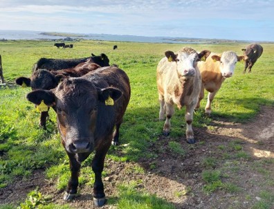
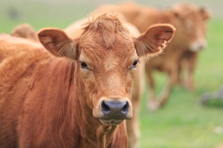
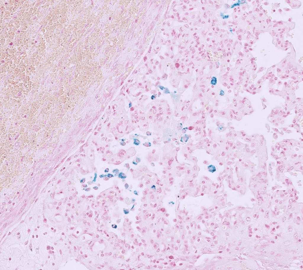
We have had a couple of submissions with positive liver fluke results in this year’s lambs in either the coproantigen ELISA or on serology testing (in Dorset & Cheshire). We have also had positive liver fluke results in these tests and on microscopy in older sheep and cattle across the country but there is always a risk in older animals that they could be carry over infections. This is earlier in the season than we would usually be expecting evidence of infection and it is likely to be as result of the wetter summer. Please note that serology on bloods or the coproantigen ELISA on faeces are more sensitive than microscopy, particularly when screening in the autumn and early winter.

BVD Cymru accredited lab.

We are an accredited lab for the Welsh BVD eradication programme. BVD antibody and antigen results will be uploaded if samples are submitted on a BVD Cymru form. As was the case with BVD Free England there is a small charge for the uploading of the results of 50p per sample for BVD antibody testing and 25p for a BVD antigen test.

Animal Health & Welfare Pathway – follow up endemic disease testing.

Please note that although we are not participating in the worm egg counting part of the AHWP for sheep we are a UKAS accredited lab and provide ISO17025 accredited tests so can be used for follow up endemic disease testing for both cattle and sheep. The diseases and conditions to be sampled for sheep include: Border disease (BD), caseous lymphadenitis (CLA), enzootic abortion of ewes (EAE), Johne’s disease, Maedi Visna (MV), toxoplasmosis, tick-borne fever, pulpy kidney, lamb dysentery, ewe nutrition status, lamb nutrition status & trace elements.

Change of bulk milk IBR ELISA test (NB IBRgE milk serology NOT affected).

Due to the IBR milk ELISA that we currently use being discontinued, we have had to switch to another supplier’s ELISA. We have trialled a couple of ELISAs from reputable suppliers whom we already work with. Both of these tests have increased sensitivity but this means that herds with positive results are now often giving results at the upper limit of the test, which is not useful for monitoring herds over time. We have therefore modified the procedure for the most suitable of these ELISAs, and carried out in-house validation, so that it can be used for monitoring of trends. Please note that the result figure from IBR milk serology cannot be directly compared to results from prior to July, due to it being a different ELISA kit, however the classification of the herd should be similar to as before. “Doubtful” results will now be classed as “suspect” as it appears likely that there is a low seroprevalence to IBR virus in these herds. Please note that the IBRgE milk serology test is not affected by this change.

New multiplex PCR test for investigating ruminant abortions.

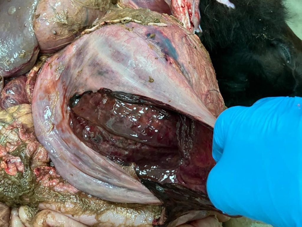
The diagnostic rate for bovine abortions through veterinary laboratories has always been relatively low. There can be a number of reasons for this – the cost of testing for the large range of possible causes makes it cost prohibitive to do a comprehensive screen, the degree of autolysis, lack of availability/testing of placenta and of course some will have been aborted for non-infectious reasons e.g. nutritional, physical or genetic reasons. With sheep abortions the diagnostic rate is higher but screening is usually limited to the most common causes. In an attempt to increase the diagnostic rate of infectious causes of ruminant abortions we are introducing a ten agent multiplex PCR test. It will screen for the following agents: Anaplasma phagocytophilum (TBF), Campylobacter fetus, Chlamydophila spp. (includes EAE), Coxiella burnetii (Q fever), Leptospira spp., Listeria monocytogenes, Neospora caninum, Salmonella spp., Toxoplasma gondii & Brucella sp. These agents are often found systemically in a foetus but can vary as to which viscera they are found in and also the levels at each site. Placenta is often a very good sample to include in a pool of tissues however if it is contaminated with faeces this can be detrimental. Faeces can contain Campylobacter fetus and also Chlamydia pecorum (a Chlamydophila sp. that does not cause abortions) which could lead to a mis-diagnosis. If placenta is faecally contaminated then it is better to sample tissues, using clean instruments and gloves, from within the carcase. The following fresh tissues, which we will test as a pooled sample, are likely to give a very good chance of detecting the above agents if they are present:
Brain (or uncontaminated placenta) & liver & foetal stomach contents (FSC)
Or if no foetus available:
Vaginal swab (plain) & uncontaminated placenta
Clean the vulva with paper towel and part the vulval lips to avoid faecal contamination of the swab and take the sample from the ventral aspect of the cranial vagina. Please note that if only a vaginal swab is submitted (taken up to five days after parturition) and no placenta is sampled then testing is unlikely to detect the presence of Neospora, Toxoplasma and probably also TBF – though serology could be done for these agents instead.
The screen does not include BVD virus (foetal fluid can be tested for BVD antigen and antibodies or there is the more expensive PCR test using fresh spleen or liver) or IBR (we have a PCR test using fresh liver, as an add on).
Please note that the screen also includes Brucella species so if a positive result is obtained for this APHA would need to be informed, with it being a notifiable disease.

Still time for flocks to gain MV Monitored-Free status in time for the sales.

  • Provides a source of low MV risk replacements especially for larger, commercial type flocks
  • Allows flocks that cannot meet the minimum two metre biosecurity boundary of the MV Accreditation Scheme to test and gain a MV status
  • Reduces the risk of a flock becoming MV infected (as bringing in untested sheep is the main route of entry for MV infection)
  • Allows low cost monitoring of flocks and will detect early entry of MV infection long before the impacts of disease occur
  • Useful for screening a non-MV accredited, commercial type flock on the same holding as an MV accredited flock
  • Infected flocks can join the scheme to benefit from reduced testing rates and to access advice from a vet with experience in the control and eradication of MV

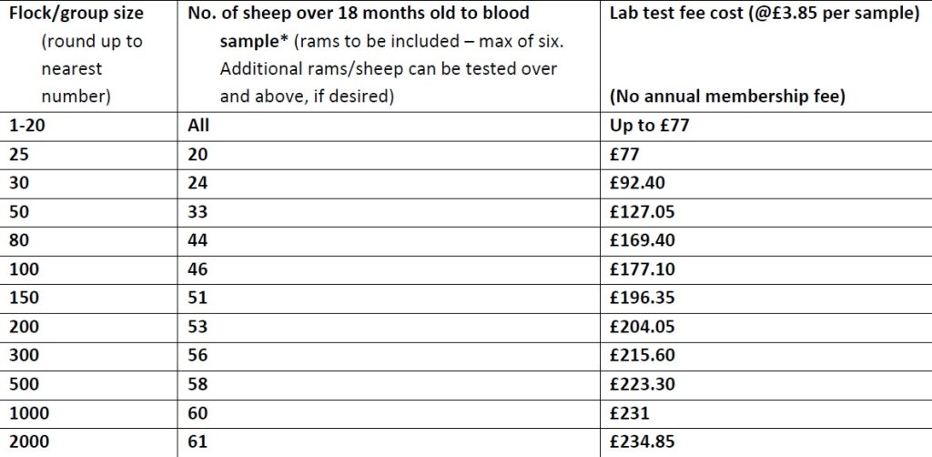
Testing of a proportion of the flock is carried out on an annual basis. Where flocks have a minimum two metre biosecure gap at all places between their flock and any neighbouring sheep they can opt to test every two years.

*Provides 95% confidence that flock infection level is below 5%

Discounted test rates for Johne’s disease & Neospora herd screens or regular batch testing.

Our Johne’s & Neospora Monitoring programmes give farmers access to discounted test rates for whole herd or regular batch testing. There is no membership fee and no set rules to follow. Johne’s serology is from £4.25 per sample and Neospora serology costs from £5.90 per sample. It works out cheaper than testing through a CHECS cattle health scheme so is ideal for herds that are testing for disease control and management purposes. Advice is provided in the lab report and farmers can be e-mailed a copy if required. Our turnaround times are very fast– often same day but within three working days for both tests. Batch testing herds also get their results in a cumulative spreadsheet. Contact us for more information at dsfarm@axiomvetlab.co.uk or on 01626 357776.
In order to avoid any unnecessary confusion, please can we ask that submission forms are only sent in with the samples and not in advance of the samples. Thank you for your cooperation.

Did you know that you can email us any additional test requests for your submissions?

You may find this is a more efficient way of making requests than phoning the farm team, saving you time in your busy day. Our farm team also find it a more efficient way of dealing with your requests.
The email address for test requests is: dsfarm@axiomvetlab.co.uk

Would you like to send the Axiom farm vets photos of PMEs to allow us to assist with sample selection and diagnosis? Please send photos by WhatsApp to 07944 649222.

Please provide your name, practice, the farmer and farm names so that we can link the photos to the relevant submission and please also indicate which Axiom vet you discussed the case with. We may wish to use some of the photos in our newsletter so please indicate if you are not happy for this to be done. All cases are anonymised and credited to the submitting vet.

Interesting cases in July


Cattle


Abortions, stillbirths, infertility, mastitis, metritis.

  • Aspergillus terreus was isolated from the stomach contents of a foetus aborted in a dairy herd. This is a known cause of mycotic placentitis and abortion in cattle.
  •  Campylobacter fetus was isolated from the stomach contents of a foetus aborted by a six-year-old suckler cow three weeks before calving.
  • Salmonella Dublin was isolated from the stomach contents of aborted fetuses in three herds. Reported clinical signs, apart from abortions, included sickness, pyrexia, retained foetal membranes and scour.
  • Neospora was detected by PCR in foetuses aborted in a number of herds, confirming infection. Histopathology of foetal heart and ideally foetal brain is needed to confirm neosporosis as the cause of abortion.
  • At least a further three cases of Q fever were confirmed this month, on PCR testing of placenta or foetal fluid. Two of the cases were from a herd with a known history of Coxiella burnettii infection. There is a statutory duty to report positive results to the APHA. Q fever is endemic in GB (approximately 80% of dairy herds are positive on bulk milk serology) and confirmation of its involvement in abortion cases requires histopathology.
  • A Welsh Black herd experienced multiple stillbirths, abortions and barren cows. The herd grazed on a mountain in the summer months and multiple ticks had been seen on the cows. All three cows sampled and tested had evidence of exposure to Anaplasma phagocytophilum, the causative agent of Tick Borne Fever, on serology, one of which had a particularly high titre suggesting relatively recent exposure. In addition, pooled plasma iodine run on six cows was low, suggesting low recent iodine intake, and the six animals seroconverted to Schmallenberg virus.
  • Histopathology of placenta from a Holstein-Friesian cow that aborted her calf six weeks before term revealed a chronic suppurative placentitis, probably due to a bacterial infection. This could have been primary, or may have been secondary to another underlying disease process.
  • Corynebacterium ulcerans was isolated from a case of bovine mastitis. C. ulcerans is an uncommon cause of mastitis in cattle. It has been reported as a zoonosis associated with consumption of raw dairy products. The zoonotic potential of different strains is due to production of a diphtheria toxin and, in this case, the isolate was confirmed as being toxigenic by the National Reference Laboratory. This is a reportable finding and the UK Health Security Agency was notified.
  • Candida krusei was isolated from a case of mastitis in a dairy cow; the cow had a hard inflamed udder and recently there had been several cows with high cell counts in the milking herd. Yeast species (such as Candida) often gain entry to the udder as a result of unhygienic intramammary infusion technique. Sometimes they may act as a true environmental pathogen when present in large numbers in the environment. Treatment with antimicrobials is totally ineffective.

Respiratory disease

  • At least eight cases of lungworm were confirmed on Baermann’s testing this month. In one case, a group of 60 five to six-month-old calves were looking very lethargic with reduced appetites. Some calves had their necks stretched out, were open mouthed breathing with some associated frothing. The animals had normal rectal temperatures but increased lung sounds on auscultation. Two tested animals were both positive on Baermann’s. A four-year-old Limousin cross cow with severe respiratory signs and two store cattle in a different herd were seropositive for lungworm.
  • IBR was detected by PCR on at least three occasions this month. In one case, histopathology of lung from one of a number of 10-month-old pyrexic calves with nasal discharge, detected bronchiolitis fibrosa obliterans consistent with a severe necrotising bronchiolitis, which in calves is often associated with a pneumotropic viral infection. Another case was suspected in a nine-month-old calf, in a herd with a history of intractable pneumonia cases, with the detection on histopathology of severe, fibrinonecrotising and suppurative tracheitis and bronchointerstitial pneumonia, which were highly suggestive of IBR.
Fibrinosuppurative inflammation lining the mucosa of the trachea suggesting possible IBR
  • At least two cases of RSV infection were confirmed by PCR this month. In one case, two calves were lost over a few weeks in a group of three to four-month-old calves on a calf rearing unit and three other animals were showing signs of respiratory disease with no response to tulathromycin. The calves had received an intranasal vaccine on arrival and an injectable vaccine three weeks after arrival, both including RSV.
  • In an ongoing animal welfare case, an animal was euthanased due to its poor condition, open mouth breathing and severe respiratory distress. Post mortem revealed more than 50% of the lung consolidated, with microabscesses throughout the consolidated areas. Histopathology was consistent with a chronic bronchopneumonia with bronchiectasis. This condition typically develops in cranial ventral lung lobes following infection with Mycoplasma bovis and subsequent infection by secondary bacterial agents (Pasteurella multocida was isolated from lung). Bronchi and bronchioles progressively dilate and fill up with mucoid exudate. In this case, there was also material including rumen protozoa in the URT suggesting aspiration of rumen contents most likely secondary to the respiratory distress described.
  • Histopathology of lung from a cow with suspected heart failure detected severe, extensive, subacute pulmonary haemorrhage with oedema and, in the pulmonary arteries, moderate, chronic arterial mural hypertrophy and hyperplasia with adventitial fibrosis. The changes in the blood vessels suggested pulmonary hypertension. Although left heart failure could be responsible for the hypertension, this is relatively uncommon in cattle and a pulmonary embolism was suggested as a more likely pathogenesis.
Macrophages containing haemosiderin (which stains blue with Perl’s Prussian Blue special stain; “heart failure cells”) in the lung of a cow.

Gastrointestinal disease

  •  Coccidiosis was confirmed with the finding of pathogenic Eimeria species (E. alabamensis, E. bovis and E. zuernii) in calves aged between three and 11 months old, where age was reported, with reported signs including malaise and scour, which was sometimes haemorrhagic in nature. Coccidiosis was also suspected in younger calves with consistent clinical signs and a high coccidial oocyst count but where speciation was not carried out.
  • A three-week-old calf died suddenly and gross examination revealed moderate ascites, extensive fibrinous peritonitis and haemorrhagic abomasal mucosa. Histopathology found tentative Sarcina-type bacteria adhered to the serosa of the small intestine suggesting leaked abomasal contents following an abomasal rupture. Perforating abomasal ulcers are a common cause of generalised peritonitis in calves. Sarcina bacteria are thought to indicate dysbiosis and are often found associated with abomasal bloat and abomasitis in young calves, where the aetiology is still not fully understood. A number of predisposing factors are thought to be involved including compromised immunity from failure of passive transfer, stress, bacterial infection of the abomasal wall, and vitamin and mineral deficiencies. The role of bacteria, such as Clostridium perfringens, C. fallax, C. sordellii and Sarcina spp., in these cases is unclear, but mixed infections have been associated with disease. Entry and proliferation of the bacteria are likely associated with damage to the abomasal mucosa. In artificial milk fed calves, this is most likely caused by poor milk hygiene, poor quality milk, large volumes of milk fed or milk fed at the wrong temperature.
Haemorrhagic abomasal mucosa in a calf with a suspected abomasal rupture and peritonitis (photo: Huw Jones, Honddu Vet Practice)
  • High pepsinogen levels consistent with parasitic abomasitis were seen in a number of ill thrifty animals. In one case, a suckler cow that was negative on faecal PCR for Johne’s in mid-June and in which there had been no improvement despite treatment for rumen fluke, the animal had loose bubbly scour for four months and was very tucked up, despite being six months in calf.
     In July, a total of 360 submissions for Johne’s disease serology and faecal PCR testing from cattle were received, with 119 submissions, including a total of 189 individual samples, testing positive. Cases included a recently bought in 15-month-old store heifer that was thin, tucked up and slightly scoured and a 26-month-old Welsh Black bull that was scouring and had reduced rumen fill.
  • Salmonella Dublin was isolated from gastrointestinal contents of two calves and faeces of one calf in a group of four to five-month-old British Friesian cross calves in which four out of 16 animals had died after a six-week period of ill thrift despite treatment for coccidiosis two weeks earlier. One of the calves also had evidence of pneumonia on PME. Salmonella Mbandaka was isolated from the faeces of a Holstein-Friesian cow, one of five cows with anorexia and increased respiratory effort, although the animals were not pyrexic and lungs were clear on auscultation. The faeces were foul smelling but there was no scour. It was also isolated from faeces samples from one of eight animals with presumed scour in a second herd. This serotype is typically feed related. Salmonella Typhimurium was isolated from a faeces sample from a two-month-old Aberdeen Angus calf with presumed scour and from a freshly calved dairy cow with cold extremities, weak rumenal contractions and milk drop. Salmonella Orion was isolated from a faeces sample from a cow with presumed scour; this isolate is more usually isolated from poultry.

Skin and eye disease

  • Chorioptes bovis infection was confirmed on microscopy of skin scrapes from an 18-month-old Limousin x British Blue heifer with a scabby area on the udder in the centre of the four teats.
  • Hepatogenous photosensitisation was suspected to be the cause of nasal ulceration in an 11-year-old Limousin cross cow that was in poor condition with a poor coat, and was scouring. GGT was elevated at 269 U/l, potential causes including fasciolosis and toxic plants, e.g. bog asphodel.
  • Moraxella bovoculi was isolated from ocular swabs taken from cattle with ocular disease in two herds. This isolate has been associated with ocular infections in some cattle but has also been isolated from the eyes of healthy cattle. It’s precise role in the pathogenesis of ocular infections is therefore not clear but it may play a synergistic role in mixed infections.

Systemic and miscellaneous disease

  •  A total of 375 submissions were received for BVD antigen testing in cattle this month, with 28 submissions, including a total of 30 individual samples, testing positive. Cases included a Holstein cross calf born with tremors and a yearling Simmental with severe scour and purulent nasal discharge, consistent with mucosal disease.
  • At least two cases of MCF were confirmed on PCR testing this month. In the first case, a one-year-old Limousin cross animal grazed with sheep, signs included bilateral corneal oedema, widespread acute skin disease and pyrexia (40.9oC). The second case, a suckler animal, also developed corneal oedema and had oral ulcers and diarrhoea. A pyrexic cow with conjunctivitis and milk drop seroconverted to MCF. This is supportive of a diagnosis of MCF but insufficient to be diagnostic as cattle can have antibodies from previous subclinical infections or from cross reactions with other herpes viruses. Heparin blood for virus PCR testing is required for a definitive diagnosis.
  • Babesiosis was diagnosed by PCR on an EDTA sample from a Shorthorn in North Yorkshire; the clinical history was not provided.
Fibrinosuppurative inflammation lining the mucosa of the trachea suggesting possible IBR

Sheep and goats

Mastitis

  • Milk samples were submitted from five ewes from the same flock which were suffering an excessive number of mastitis cases in comparison to previous years. Pure and heavy growths of Staphylococcus aureus were cultured from two of the samples. Mannheimia haemolytica was isolated from two further samples in heavy growths and in the fifth sample the predominant growth was Streptococcus uberis with Mannheimia haemolytica also present in moderate growth. In a further case of mastitis in a flock of Beulah speckled ewes, which were reported to have necrosis of the udder and some ewes had died, Staphylococcus aureus was isolated. Mannheimia haemolytica and Staphylococcus aureus are the most commonly isolated bacteria in cases of mastitis in sheep and outbreaks can be associated with poor milk supply in ewes resulting in over vigorous suckling by lambs and trauma followed by invasion of bacteria.

Respiratory disease

  • A marked, extensive, subacute, necrosuppurative and fibrinous bronchopneumonia was the cause of acute deaths in lambs in one flock. From the changes observed on histopathology, the bronchopneumonia was thought most likely due to a Pasteurellaceae with Mannheimia haemolytica being considered the most likely pathogen.
  • Atypical pneumonia was identified by histopathology in a yearling sheep which was one of six to have died suddenly. At post mortem examination, the lungs were noted to be dark red/ purple in colour. Only one injection of a course of multivalent clostridial/pasteurellosis vaccine had been administered to the group. Atypical/ enzootic pneumonia in sheep has a multifactorial aetiology including Pasteurella multocida, Mannheimia haemolytica, Bibersteinia trehalosi, staphylococci, streptococci and Mycoplasma ovipneumoniae. Lambs normally present with ill thrift and a mild cough. In this case, secondary bacterial pneumonia was identified which could account for the sudden death.
  • Ovine Pulmonary Adenocarcinoma was diagnosed on histopathology of lung from ewes in two flocks. One case had gross evidence of pleurisy and pericarditis with mild and chronic secondary bacterial infection observed microscopically.
  • Streptococcus pyogenes was isolated from swabs of a lung abscess taken at post mortem of an adult ewe. The vet reported that there had been seven ewe deaths in the space of a week. The post mortem revealed one large abscess in the right caudal lung and smaller abscesses throughout the lung tissue including some consolidated areas. S. pyogenes (Group A Streptococcus) is not part of the normal bacterial flora of animals and is not a common environmental contaminant. It was thought to be a significant finding in this case. Histopathology identified a chronic purulent bronchitis/ bronchiolitis and in the sections examined there was no sign of underlying OPA or other predisposing disease.
  • The chest cavity of a two-year-old Bluefaced Leicester ram was noted to be ‘full of masses’ at post mortem and a sample of the presumed tumours were submitted for histopathology. A densely cellular, unencapsulated proliferation of neoplastic round cells forming closely packed sheets were observed. This appearance was typical of a round cell tumour, most likely lymphoma. A thymic lymphoma or possibly a multi-centric lymphoma (if other masses were present elsewhere) was considered most likely. Lymphoma is one of the more common neoplasms in sheep, often seen as sporadic cases in older animals. As seen in cattle though, sporadic and enzootic forms of lymphoma can also be seen in sheep and it was noted that it is not possible to rule out infection following exposure to retrovirus (Bovine Leukaemia Virus (BLV) by histopathology alone.
Blue neoplastic cells effacing a large portion of lung in a sheep with likely lymphoma

Gastrointestinal disease

  • In July, a total of 79 submissions for Johne’s disease serology and faecal PCR testing from sheep and goats were received, with 17 submissions, including a total of 27 individual samples, testing positive. In one flock, two-to
    three-year-old ewes were observed to be wasting with a dark scour present shortly before death. In the last year, 12 of the flock of 300 had died with similar signs. An individual tested as seropositive for Johne’s but also had a strongyle egg count of 5500epg indicating parasitic gastroenteritis and possible haemonchosis was also present and this could have been the main cause of the clinical signs. A two-year-old male goat which had weight loss, anaemia, hypoalbuminaemia weakness and inappetance was seropositive for Johne’s disease. In a post mortem case of a ewe with bottle jaw and excess fluid in the abdominal cavity, a sample of faeces tested positive for Mycobacterium avium paratuberculosis (MAP) with a low Ct value of 20 indicating heavy shedding of MAP. There was a high strongyle egg count in that case too with a count of 1400 strongyle epg.
  • There was evidence of fascioliasis in a group of young ewes of different breeds of which only the Suffolk ewes were reported to be affected by scour and malaise. Fluke eggs were observed on microscopy and there was a significant high strongyle egg count of 1650epg indicating concurrent parasitic gastroenteritis.
A liver fluke in the liver of a sheep
  • Haemonchosis and nematodirosis were diagnosed in a 12-week-old Texel cross lamb which had presented as a sudden death. There were adult nematodes present in the abomasum and the carcase was pale. The faecal worm egg count was 8500epg strongyle species and 500epg Nematodirus battus. Of the strongyle eggs present, 99% were identified as Haemonchus species. On another holding, a three to four-month-old lamb had a worm egg count of 7170epg and had signs of scour. In that animal, 42% of the 7000 strongyle epg were Haemonchus sp.. In a third case, sheep had pale mucous membranes and two deaths had occurred. Of the strongyle count of 660epg only 8% (approximately 50epg) were identified as Haemonchus species. However, it was noted that Haemonchus burdens can vary significantly within flocks and, with evidence of a significant parasite burden, anthelmintic treatment was warranted. There were at least three more sheep flocks this month where Haemonchus infection was confirmed and several in which haemonchosis was strongly suspected. One sample from a sick Valais Blacknose lamb had a strongyle count of >50,000epg.
  • Anthelmintic treatment failure was suspected in a group of ewes and lambs which had had treatment for Haemonchus two weeks previously using closantel. Eight groups were sampled and strongyle egg counts varied from 400epg up to 3750epg, a high count still suspicious of the presence of Haemonchus. A group of four to five-month-old lambs had samples taken 14 days after a clear (avermectin) drench. The strongyle egg count had previously been 1550epg and post treatment the count was 450epg suggesting partial treatment failure. A goat which was reported to have a persistently high worm egg count was sampled 14 days post dosing with a benzimidazole wormer; there was a strongyle egg count in the sample of 750epg.
  • Coccidiosis was identified in weaned lambs on at least two holdings this month. In the first case, the coccidial oocyst count was 25,900opg of which 29% were of the pathogenic species Eimeria ovinoidalis. On the second farm, there was a coccidial oocyst count in scouring lambs of 12,000opg and of these 36% were identified as E. ovinoidalis and 1% were of the other pathogenic species in sheep, E. crandallis. In a group of 11 10-week-old scouring goat kids, some of which were reported to be lethargic and tucked up, there was a coccidial oocyst count of >50,000opg of which 80% of the Eimeria species were of the pathogenic species, E. arloingi, 10% E. christenseni, 2% E. ninakohlyakimovae and 2% E. hirci.
  • Type D clostridial enterotoxaemia was the suspected cause of death in an 18-month-old male Golden Guernsey goat that died after an acute episode of diarrhoea. On PME, the small intestinal mucosa was haemorrhagic, the contents were very watery and C. perfringens alpha and epsilon toxin were detected in caecal contents. The sheep adapted strain of Salmonella enterica ssp. diarizonae, Salmonella 61:k:1,5,7, was also isolated.

Skin and eye disease

  • Positive antibody titres to Corynebacterium pseudotuberculosis (CLA) were detected in a three-month-old goat kid with a facial abscess and a Charollais shearling tup with an abscess in the region of the submandibular lymph nodes.
  • Orf (parapox) virus was detected by PCR testing of skin scabs from a two-year-old Leicester ram with proliferative papillomatous lesions on the head.
  • Psoroptes ovis mites were observed on at least two occasions, from a four-year-old Texel ram who was pruritic over the back and also lethargic and depressed, and from a group of lambs that were pruritic over the back and sides of the body. Antibodies to Psoroptes ovis mites were detected in at least a further four flocks in which signs of sheep scab had been observed.
  • Moraxella bovoculi was isolated from an eye swab from a lamb with signs of keratoconjunctivitis. This organism can be found in cases of infectious keratoconjunctivitis but it can also be found in healthy eyes. It is thought that it may not be a primary agent but may act with other agents to cause infection. The commonest cause of pinkeye in sheep is Mycoplasma conjunctivae, for which a PCR test is available (plain swabs from up to three sheep, if pooling required).
  • Mycoplasma conjunctivae was detected in eye swabs from a goat flock with persistent pinkeye issues. In this case, Pseudomonas sp. were also isolated and this could have been acting as an opportunistic pathogen. In a case of eye infections in a sheep flock, there was also evidence of selenium deficiency in the group. Low selenium status is associated with poor immune system function and resulting increased susceptibility to infections although a specific link to Infectious Keratoconjunctivitis is unproven.
  • Staphylococcus aureus and Streptococcus dysgalactiae were isolated from ulcerative lesions around the face and ears of two Texel shearlings. The lesions were reported to spread quickly once established, forming scabs and pus with hair loss over affected areas followed by gradual healing. Both of the isolates were considered to be of significance in this case of dermatitis/periorbital eczema. Trauma e.g. head rubbing/bumping if being trough fed, would be the most common underlying cause resulting in skin abrasions and allowing bacterial entry.
  • Streptococcus ovis was cultured from swabs of a facial abscess in a dairy goat. The suspected origin of infection was a molar tooth root abscess.

Systemic and miscellaneous disease

  • Cardiac failure was suspected as the possible cause of severe and chronic passive congestion identified on histopathology of liver tissue from a four-month-old female goat kid which was reported to have a large liver at PME. Although the changes in the liver were typical of those seen in cases of cardiac failure, it could not be confirmed as no gross lesions of the heart were described and no heart tissue was available for histopathological examination.
  • Maedi-Visna was identified by serology in a ram in poor condition in an upland flock, in a five-year-old ewe with evidence of carpal arthritis and in a six-year-old ewe with wasting and pale mucous membranes (concurrent haemonchosis was suspected). MV was also identified in screening tests in several flocks where no clinical signs had been observed (or no further history was supplied).
  • OvHV-2 (the causative agent in Malignant Catarrhal Fever) was detected by PCR in blood samples in all six Cameroon sheep which were part of a zoological collection. Sheep are known to carry and shed OvHV-2 without showing clinical signs except in rarely recorded cases, but the virus can cause severe disease and death when transmitted to bovine and cervine species.
  • A young ewe which was found recumbent and in poor body condition with dilated pupils, nystagmus and congested mucous membranes was positive on serology for Anaplasma phagocytophilum, the causative agent of Tick Borne Fever. Although TBF infections are often subclinical, they can predispose to severe illness and death from Louping ill virus due to immunosuppressive effects. Louping-ill testing was not carried out on this submission.
  • A mass removed from the jaw of a seven-year-old ram, which had loose molars, was found to be a poorly differentiated malignant neoplasm. A spindle cell tumour such as a fibrosarcoma or tumour originating from the bone, or an amelanotic melanoma were thought to be possible given the appearance and location of the mass.
  • Uroliths were observed on post mortem of a pet castrated male sheep which was found collapsed with abdominal distension and had not passed urine for more than 12 hours. The bladder wall was thickened and histopathology identified an acute inflammatory response, typical of what would be expected in a case of urolithiasis and urinary obstruction. Urolith analysis found the stones to be composed of 100% calcium carbonate. In one study, calcium carbonate was found to be the second most common type of urolith in sheep and goats, with the most common being amorphous magnesium calcium phosphate in combination with struvite (magnesium ammonium phosphate). Calcium carbonate calculi tend to be more common in animals on pastures rich in clover or oxalate containing plants.
  • Injection site abscesses developed in 25 to 30 of a group of 185 Easycare lambs three weeks after receiving a multivalent clostridial/Pasteurella vaccine. Some lambs had very large abscesses involving the entire neck, with large quantities of pus present. None of the animals were sick. Some abscesses were lanced and swabs from three cases were submitted for culture; in all three cases, Trueperella pyogenes was isolated, a known cause of pyogenic infections in livestock. It had started to rain heavily halfway through vaccinating.

Deficiencies and toxicities

  • Cobalt deficiency was diagnosed in sheep, goats and alpacas this month, reported clinical signs including ill thrift and skin issues. It was also seen, together with marginal selenium, in one of a group of ewes which exhibited weakness and neurological signs progressing to seizures and death, although the limited testing carried out did not rule out other potential explanations for the signs.
  • Copper deficiency was confirmed on liver biochemistry in a three-year-old Boer goat that had a history of gastrointestinal parasitism and poor body condition, but at the time of death had no premonitory clinical signs. It was also confirmed on liver biochemistry in a three-month-old beef calf in a herd in which multiple calves had died over the last few months. Low plasma copper levels were also seen on sampling animals with presenting signs including failure to lose the winter coat, coat colour changes, hair loss, ill thrift and cows producing stillborn and weakly calves.
  • Cobalt and copper deficiency were diagnosed in an anaemic, hypoalbuminemic Anglo-Nubian goat that also had a strongyle egg count of 1000 epg and collapsed when the blood sample was taken. Low copper and vitamin B12 levels were found in ill thrifty lambs, a very thin, anaemic 12-year-old goat and a reindeer that was losing condition and had loose faeces.
  • Low copper and plasma inorganic iodine levels were reported in suckler cows producing weak calves and in ill thrifty suckler calves.
  • Low plasma inorganic iodine levels were also seen in ill thrifty cattle and sheep.
  • Selenium deficiency was diagnosed particularly in cattle this month, with the predominant presenting sign being ill thrift. Other presentations included a four-month-old suckler calf going off its legs, the second to do so since the cows and calves were turned out six weeks earlier, half of a group of 16 to 20-month-old Holstein-Friesian heifers showing little ovarian activity, and Charolais cross cows in good body condition at calving whose calves were dying within a few days of birth despite receiving colostrum.
  • Hypomagnesaemia, hypocalcaemia and/or hypophosphataemia was seen in a number of recumbent cows this month.
  • Ketosis was confirmed in an intermittently recumbent Jersey cow, with loose dung and exhibiting odd behaviour, with the finding of a serum BHB result of 4.83 mmol/l.
  • Low or suboptimal urea levels were reported in a number of cattle primarily presenting with ill thrift and also, in one Holstein-Friesian herd, with reduced milk yield. Low urea indicates a lack of rumen degradable protein, caused by either inadequate levels in the diet or insufficient intake.
  • One of the cases of vitamin A deficiency seen this month was in one of three three-year-old Limousin bulls at pasture with acute onset lameness and coronary band pathology. In a second case, neonatal calves were becoming pyrexic in the first two days of life despite very good management and two calves sampled were both low for vitamin A. There was a history of vitamin A deficiency in the herd.
  • Copper toxicity was diagnosed on liver biochemistry in a male bovine that had scoured for a few days and had a very yellow liver on PME, in an 18-month-old ram that was dull, jaundiced and pale for a few days before dying and had been grazing with horses, and in one of two dead sheep with unknown history.
  • Lead toxicity was diagnosed on liver biochemistry in a pre-weaned calf in a suckler herd. The animal was found dead at grass and, when the remainder were walked in, a couple of the calves appeared blind. The level of lead in the liver also raised a food safety issue and it was advised that the APHA be contacted. A second case extended back to October 2023 when two out of 22 animals were reported dead and high lead levels were found at PME. A two-year-old Charolais cross steer/bull was retested and its blood lead level had fallen from 0.55 mmol/l in March 2024 to 0.24 mmol/l in July 2024. At this level, it is advised that animals should only be presented for sale or to the food chain with food chain information and this should state that offal should be discarded.

Camelids

Skin and eye disease

  • Staphylococcus aureus was isolated from the site of jugular catheterisation in an alpaca.
  • A sixteen-year-old male alpaca had a mass removed from the lateral hindlimb between the stifle and hock. The mass was noted to be friable and spongy in texture. Histopathology indicated that it was a sarcoma most likely a leiomyosarcoma. Tumours of the skin may originate from blood vessels or arrector pili muscles. In common with other sarcomas of soft tissue origin, these tumours tend to be locally infiltrative but are slow late to metastasise. In this case full excision could not be confirmed and therefore recurrent growth was thought to be likely.

Gastrointestinal disease

  • Parasitic gastroenteritis was thought to be the cause of illness in a 16-year old male castrate alpaca which had shown signs of colic, reduced gut sounds and diarrhoea, and was in poor body condition. There was a strongyle egg count of 500epg.
  • Coccidiosis was suspected as the possible cause of tenesmus, rectal prolapse and faecal staining of the perineum in a five-week-old cria. Although none of the oocysts observed in this case were of the most pathogenic species in camelids, E. macusaniensis, the coccidial oocyst count of 750 opg was still considered of possible significance in this age of animal.

Systemic and miscellaneous disease

  • A seven-year-old female alpaca showed signs of chronic weight loss and colic symptoms. An abdominal mass could be felt on palpation of the abdomen. At post mortem, the mass was visualised and histopathology identified a variably differentiated malignant neoplasm which was considered most likely to be a lymphoma or histiocytic sarcoma.
  • Trueperella pyogenes was isolated from a thoracic abscess identified at post mortem of an adult alpaca.

Other Species

    • An 80-week-old Dekalb hen underwent post mortem examination and it was noted there was a cystic appearance to the intestinal tract along with evidence of peritonitis. The appearance of the tissue on histopathological examination was consistent with adenocarcinoma. The tissue of origin of the neoplasia was not clear, however, the usual origin in chickens is ovarian.
    • ZN staining of the liver of an adult pheasant confirmed avian tuberculosis as the cause of a granulomatous hepatitis identified on histopathology, most likely due to Mycobacterum avium. There had been more than 100 deaths reported since moving the birds two weeks previously (the total numbers in the group were not known). M. avium is ubiquitous in the environment, and many wild birds are infected. Pheasants are particularly susceptible to infection.
    • Mycoplasma gallisepticum was detected by PCR testing of swabs from a hen with signs of swelling of the head and nasal discharge.
    • Parasitic gastroenteritis was diagnosed in a six-year-old boar with ill thrift and poor body condition which had not received any worming treatment for over two years. There was a strongyle egg count of 3550epg.
    • A 12-year-old reindeer, which was the only one of a group of three which had signs of scour, tested positive on faecal PCR for the presence of MAP (Mycobacterium avium paratuberculosis). The presence of MAP in faeces is supportive of a diagnosis of Johne’s disease.
    • Listerial encephalitis was diagnosed by histopathology of the brain in a fallow deer which was euthanised after being found unwell and circling in one direction.

Follow us on Facebook

This newsletter is sent by e-mail to each vet practice but if you would like a copy sent to your individual e-mail account please contact us at dsfarm@axiomvetlab.co.uk and we can add you to our circulation list.

Related Posts