Farm January Newsletter 2026

Dedicated farm line – 01626 357776

Follow us on Facebook

https://www.facebook.com/Axiomfarmvetlab/

This newsletter is sent by e-mail to each vet practice but if you would like a copy sent to your individual e-mail account please contact us at dsfarm@axiomvetlab.co.uk and we can add you to our circulation list.

Would you like to send the Axiom farm vets photos of PMEs to allow us to assist with sample selection and diagnosis? Please send photos by WhatsApp to 07944 649222.

You may find this is a more efficient way of making requests than phoning the farm team, saving you time in your busy day. Our farm team also find it a more efficient way of dealing with your requests.
The email address for test requests is: dsfarm@axiomvetlab.co.uk 

Blankety Blank

We love getting your samples to test, but please let us know on the form which veterinary practice they have come from! Each month we receive hundreds of submissions form with no practice details. Luckily the postal room and diagnostic support teams are great at detective work and many of the mystery samples can be traced. However, trying to track down the origin of samples can significantly delay their arrival in the testing departments, potentially increasing turnaround times. The most up to date versions of the forms can be found on our website to download but note these do not automatically include practice codes or details so these need to be added before sending.

Blankety Blank

Axiom farm vets

If you wish to speak to a particular farm vet about one of your cases, you can consult the table below to see when our vets are usually available. A duty farm veterinary advisor is always around on a Saturday morning from 9am-12pm.

Axiom farm vets

Malignant Catarrhal Fever PCR Test

We are now carrying out the PCR test for Ovine Herpes virus -2 (the causative agent of Malignant Catarrhal Fever in cattle) in-house with an improved maximum turnaround time of five days. EDTA whole blood is the preferred sample type but heparin whole blood and plain nasal swabs can also be tested. For postmortem cases a minimum of 1g of tissue (lymph node ,spleen, lung , liver or thymus) can be tested.

Haemonchus Identification

We have validated a new method for differentiation of Haemonchus species from other Strongyle eggs in faecal samples. The new technique uses digital technology (Ovacyte) to identify Haemonchus sp. The turnaround time for the test has been improved whilst maintaining comparable sensitivity and limit of detection to the peanut agglutination test. We are hopeful that this will assist vets in rapidly detecting and dealing with increasing numbers of haemonchosis cases on farms throughout the UK.

Discounted Johne’s disease & Neospora testing

Our Johne’s & Neospora Monitoring programmes give farmers access to discounted test rates for whole herd or regular batch testing. There are no membership fees and no set rules to follow. Johne’s serology is from £5 per sample and Neospora serology costs from £6.75 per sample. It works out cheaper than testing through a CHECS cattle health scheme so is ideal for herds that are testing for disease control and management purposes. Advice is provided in the lab report and farmers can be e-mailed a copy if required. Our turnaround times are very fast– often same day but within three working days for both tests. Batch testing herds also get their results in a cumulative spreadsheet. A reminder to test email is sent out for herds on annual testing. Contact us for more information at dsfarm@axiomvetlab.co.uk or on 01626 357776.

Johne’s ATV’s

What is an appropriate Johne’s ATV for dairy herds using blood testing?
The concept of a setting a target Average Test Value (ATV) is not only to encourage progress with Johne’s control but also to discourage retention of known infected animals in the herd. The Johne’s Control Initiative set an ATV target of 5.5 for herds using milk serology but it was unknown what an appropriate target should be for dairy herds using the more sensitive blood serology for screening.

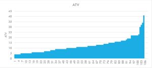
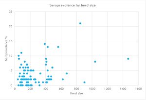
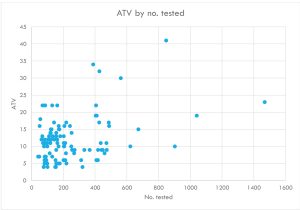
We have therefore reviewed our blood serology data for 62 dairy herds with up to four complete years of herd screening results. ATVs were calculated for 105 complete sets of adult herd tests (not including 30-60 cow subset screens). The ATVs ranged from 4 to 41:

The ATVs ranged from 4 to 41:

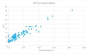
The ATVs were plotted against the herd seroprevalences and there is a good correlation:

ATV by seroprevalence

There was a tendency for larger herds to have a higher seroprevalence:

Seroprevalence by herd size

This is also reflected in the ATVs as follows:

ATV by no. tested

The ATVs were put into bands and the approximate seroprevalences are shown in the table below:

ATVs were put into bands

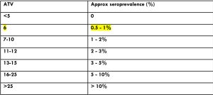
A target of below say 12 could be chosen initially, which equates to a seroprevalence of approximately 2-3%. As the herd progresses the target could be adjusted to say 6, which would be an approximate blood seroprevalence of below 1%. This ATV target of 6 on blood serology is similar to the milk target of 5.5 set by the JCI (we are unaware what milk seroprevalence their ATV relates to. Milk serology is also less sensitive than blood serology). For high seroprevalence herds their initial target could be set at perhaps 20 initially.
A herd with an ATV of 4 and no positives detected is likely to be a lower risk herd than a herd with an ATV of 10 where there had also been no positives identified as there could be a higher risk of subclinically infected animals being present in the latter.

Liver fluke – the situation so far this winter

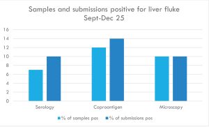
In the current autumn-winter period only relatively small numbers of submissions have tested positive for liver fluke.
Using the coproantigen ELISA on faeces 1 in 7 submissions (75 from 536) have tested as positive (14% of submissions, 12% of the individual samples (106 from 869).
About 60% of the positive submissions were from sheep and just over a third were from cattle with camelids making up the remainder.

  • Individual samples accounted for 27% of the positives.
  • Pre-pooled samples made up 49% of the positives.
  • Pools made up of 3-5 samples at the lab had 16% of the positives.

(With the remaining 8% it was unclear if they were from individuals or pre-pooled)

With the serology test one in ten submissions had positive results, indicative of exposure, and it was same level of positive submissions with fluke microscopy for eggs.

957 individual blood samples tested of which 63 were positive = 6.6% of samples

235 pooled samples were tested for antibodies of which 15 were positive = 6.4% of samples

The higher proportion of positive samples in the fluke antigen ELISA appears to be down to samples from the west of Scotland, which were mainly tested with this method and the vast majority of them were positive.

Liver Fluke Submissions

It is likely that drought conditions in some areas in recent years have eliminated or reduced the number mud snails, which are an essential part of the liver fluke life cycle. Encourage farmers to test to determine if treatment is required rather than automatically treating for liver fluke, which could save them money and time. It is of course important to sample sufficient animals.

Please use field kits with herd screens

When sending submissions containing 50+ bloods, please ensure that you use a Field Kit containing a polystyrene sample rack/box for the orderly transportation of your samples, remembering to populate the rack in the same order as your accompanying animal ID list. Receiving large quantities of blood samples in a plastic bag or cardboard box is not appropriate or conducive to the efficient handling of such submissions and invariably leads to significant delays in preparation and turnaround times.
Field Kits (filled with the required serum gel tubes) are readily available to order via the following link… https://milab.store.unleashedsoftware.com/

Interesting cases in November

Cattle
Abortions

  • Trueperella pyogenes was isolated from the stomach contents of an aborted foetus. The dam aborted the foetus at around five months gestation following signs of illness and a drop in milk yield in the preceding days.
  • Listeria monocytogenes was isolated from a bovine foetus aborted at 4-5months gestation.
  • Salmonella Dublin was identified as the cause of abortion in six cases in November and Salmonella Montevideo in two abortion cases.
  • Seroconversion to Salmonella Dublin was demonstrated by paired S.Dublin antibody ELISA testing in a Holstein-Friesian cow from a herd with higher-than-expected abortions and early embryonic losses. Of three other cows from the herd which were tested on the same submission (single serology) two were positive for antibodies to Salmonella Dublin.
  • Q fever (Coxiella burnetii) was detected by PCR in one abortion case in November. Detection of this agent in PCR could be an incidental finding as histopathology is required to reach a diagnosis.

Respiratory disease

  • Haemothorax, lung abscessation and consolidation of lung tissue were observed at postmortem examination of an adult dairy cow. Microscopically, pulmonary vascular abscess formation with accompanying fibrinous pneumonia were observed (Figure 1). The findings supported the practitioners’ suspicion of pulmonary thromboembolism as the cause of death in this case. Pulmonary thromboembolism in cattle often arises secondary to rumenitis and liver abscessation leading to thrombus formation in the caudal vena cava.

Cow pulmonary thromboembolism.

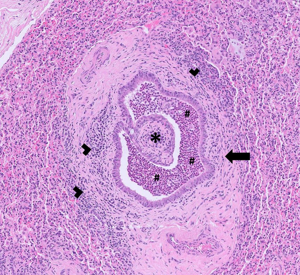
Figure 1: Cow pulmonary thromboembolism. Caption ‘A large vessel occluded by fibrin, inflammatory cells and bacteria in a cow with a septic pulmonary thromboembolism’.

  • A dairy farm reported issues with heifers joining the milking herd which then developed nasal discharge, followed by chronic mucoid discharge. Paired blood samples were taken from acute cases to check for seroconversion to respiratory pathogens. Of five animals sampled one had clear evidence of seroconversion to BVD between first and second samples. Of the others two had high antibody levels which remained high on the second sample, one went from a low positive to a high positive result and the remaining animal appeared to be in the process of seroconversion to BVD having gone from a negative to a suspicious result for antibody to BVD. One of the five animals also seroconverted to PI3 virus.
  • Salmonella Dublin was isolated from pooled nasal swabs of neonatal (48hrs old) dairy calves showing signs of pyrexia and respiratory disease. Freshly calved cows were also showing signs of pyrexia. One recently calved cow which died following a short illness and was sent to a postmortem facility was diagnosed as having salmonellosis caused by Salmonella Dublin. Salmonellosis can occasionally present as neonatal calf illnesses or deaths/ illness in adult cattle with out obvious signs of enteritis. We can also test nasal swabs in the Salmonella PCR test if there are concerns over its involvement in respiratory disease.
  • Postmortem tissues were received from a neonatal beef calf (six hours old). It was the third calf to die of a similar age in the herd. Histopathology identified a moderate to marked, necrotising interstitial pneumonia with lymphocytic bronchiolitis. The lymphoplasmacytic infiltrate suggested an insult of longer duration than the age of the calf, suggesting a likely in-utero insult (e.g. a chronic bacterial infection). The necrosis of terminal airways and interstitium was indicative of a more acute change. Possible pathogens which could be responsible for such changes include BHV-1 IIBR) , RSV, PI3, Mycoplasma bovis, Ureaplasma diversum, and BVD. It was considered possible that a placentitis could have been present and if there were further cases histopathology of the placenta was recommended.

Gastrointestinal disease

  • An adult Longhorn cow died after showing signs suspicious of hepatic encephalopathy. There was a history of liver fluke infestation in the herd. Histopathology identified severe and widespread hepatic fibrosis which would likely be associated with hepatic failure. Hepatic encephalopathy in cattle often presents as reduced appetite and depression and may progress to head pressing, visual impairment, circling, seizure and coma or can even present as sudden death. It is thought that the neurological signs may be related to accumulation of ammonia and by-products of amino acids in the brain secondary to liver dysfunction. Cirrhosis due to long term damage by liver fluke was thought to be the possible underlying cause in this case though toxins from plants, particularly those containing pyrrolizidine alkaloids (e.g ragwort), endotoxaemia (eg due to mastitis, metritis) and fatty liver disease are all possible causes.
  • E.coli F5 (K99) antigen was detected in faecal samples from a neonatal calf. The presence of F5 antigen in neonates is consistent with enterotoxigenic E.coli infection. Colonisation of the intestinal tract by the bacteria causes excessive secretion of fluid into the intestinal lumen and rapid dehydration. After five days of age the F5 antigen can no longer bind to the intestinal mucosa and calves are no longer susceptible to this infection.
  • Salmonella enteritica subsp. enterica serotype Stanleyville was isolated from faecal samples from a ten-week-old dairy calf with scour. Salmonella Stanleyville is a rare serotype in the UK having been isolated only once from horses and once from poultry since 2021.
  • A recently calved heifer became ill and recumbent with profuse scour. PCR testing for MAP was positive with a Ct value of 22 indicating heavy shedding of MAP bacteria consistent with diagnosis of Johne’s disease. Liver and rumen fluke eggs were seen by microscopy confirming patent fascioliasis and patent rumen fluke infection.

Skin and eye disease

  • A Rothia sp. was isolated from eye swabs from a Limousin cross cow which was one of numerous cattle in the herd with signs of recurrent Infectious Keratoconjunctivitis. The problem had started during the summer, but more new cases had occurred since housing. Mixed bacterial growths of mainly commensals and contaminants were cultured from two eye swabs but in one of these a Rothia sp. was identified. Rothia sp. have been found with higher frequency in diseased eyes than in healthy eyes in one study, though the significance of the finding was uncertain.

Systemic and miscellaneous disease

  • A group of spring born dairy cross calves presented with lameness, inflammation of the interdigital space and pyrexia of >400C. Large numbers of ticks were seen on the animals at clinical examination. EDTA blood samples were taken and tested for the presence of Anaplasma phagocytophilum by PCR. Two of three animals tested were positive confirming Tick-borne Fever. Although Foul-in-the-foot or interdigital necrobacillosis, caused by Fusobacterium necrophorum was the suspected cause of the foot lesions in this case, this condition is not specifically associated with Tick-borne-fever. TBF is however known to cause immunosuppression which can predispose to bacterial infections.
  • Blood samples were received from a cow in county Somerset which was reported to be pale and lethargic. Haematology revealed significant anaemia with a PCV of 11% (RI 24-40%) and an absolute reticulocyte count of 34x 10^9/L indicative of a regenerative response. Reactive lymphocytes were observed consistent with antigenic stimulation. PCR testing for Babesia sp. was positive confirming a diagnosis of Babesiosis. Magnesium was 0.5mmol/l (RI 0.7-1.3mmol/l) indicating there was also hypomagnesaemia.
  • A ten-year-old Belted Galloway cow which had recently dramatically lost condition and had a dry cough tested positive for BVD virus in the antigen ELISA with a result indicative of a high level of virus (3.9OD). Optical Density (OD) is a measure of how much light is absorbed by a sample in a spectrophotometer. In the context of ELISA, it reflects the amount of colour developed in the wells of the assay plate, which is proportional to the concentration of the target analyte (e.g proteins antibodies) present in the sample.

Sheep and goats

Respiratory disease

  • Postmortem tissues were received from a four-year-old Alpine nanny goat. There had been ten deaths in the herd over only three days. The goat which was examined had shown respiratory signs with a mucoid discharge at the nostrils. Congestion of the lungs was apparent on gross examination leading the submitting veterinary surgeon to suspect systemic pasteurellosis as the cause of death. The predominant changes in the sections of lung examined included severely distended peribronchiolar glands filled with fluid, dilated airways, atelectatic alveoli and pulmonary oedema. These changes were considered consistent with alveolar proteinosis (an abnormal accumulation of proteins and fats within alveoli), which in goats is most often caused by CAE lentivirus. However, it was noted that a diagnosis of CAE was unlikely to explain the recent high mortality as it would be unusual for several animals to reach a stage of severe terminal disease around the same time, unless perhaps there was a particular stressor or environmental changes that could have exacerbated pre-existing disease.
  • In one sheep flock there were two ewe deaths a fortnight after anthelmintic treatment for high gastrointestinal parasite burdens. Focal solid lung lesions were observed at postmortem leading to suspicion of ovine pulmonary adenocarcinoma (OPA). However, histopathology findings were not supportive of a diagnosis of OPA. Instead, it was noted that the pulmonary parenchyma was replaced multifocally by nodular areas of fibrosis in which numerous embedded helminth parasites, present as eggs, larvae, and adult worms, associated with variable inflammation including eosinophils, multinucleate giant cells and macrophages. Adult worms could be seen within airways, sometimes with rupture of the airway and sometimes with mild airway epithelial hyperplasia. In some of the areas of fibrosis the residual alveolar spaces were lined by type II pneumocytes (reactive type II pneumocyte hyperplasia). Variable numbers of lymphocytes and plasma cells were also present within the areas of fibrosis. Verminous pneumonia was confirmed and the presence of adult worms within the airways suggested involvement of Dictyocaulus filaria. Clinical disease due to lungworms is less common in sheep than in cattle though coughing and ill-thrift are reported in animals with heavy burdens. There was no evidence that the lungworm infestation was related to the ewe’s death in this case.
  • Salmonella 61:k:1,5,7 was isolated from nasal swabs taken from sheep. The flock had ongoing issues with coughing and nasal discharge in the sheep. This Salmonella serovar is a host adapted strain of Salmonella sp. and has been associated with chronic proliferative rhinitis (CPR). The disease causes a proliferative inflammation of the nasal turbinates causing obstruction of the nasal cavity, dyspnoea and nasal discharge. CPR cases could be a risk for spreading Salmonella 61:k:1,5,7 to other animals/flocks as well as having zoonotic potential.

Skin and eye disease

  • CLA (caseous lymphadentitis) infection was diagnosed by serology in two sheep on different holdings, in a Cheviot tup with a chronic submandibular mass and a Kerry Hill ewe with scarring over the parotid lymph nodes. There was one further case in which Corynebacterium pseudotuberculosis was cultured from an abscess at the base of a sheep’s ear.

Systemic and miscellaneous disease

  • Pseudomonas aeruginosa was cultured from vaginal swabs from two ewes which had been flushed for embryo transfer and were showing signs of endometritis.

Camelids

Gastrointestinal disease

  • Tissues were received from a PME carried out on a female alpaca by Veterinary Investigation Services Ltd which had detected megaoesophagus, severe chronic mange and aspiration pneumonia. Histopathological evidence of oesophageal muscle degeneration supported the diagnosis of megaoesophagus. Megaoesophagus is typically an idiopathic condition in camelids, but possible causes in other species that could be considered include iron deficiency, lead toxicity, organophosphate toxicity, toxoplasmosis and white muscle disease (Watrous et al, 1995, JVIM 9: 92-99). Ectoparasites were found in sections of haired skin with hyperkeratosis and aspiration pneumonia was also confirmed.
  • Parasitic gastroenteritis was diagnosed in Bactrian camels in two different collections, with high strongyle egg counts and, in some samples, the presence of Trichuris eggs which are known to be pathogenic in camelids, unlike domestic ruminants.
  • Fasciolosis was confirmed in a llama on coproantigen ELISA testing. This has proved to be a useful diagnostic test for fasciolosis in camelids.

Systemic and miscellaneous disease

  • An ulcerated nodule on the inside of the lower lip of a 13-year-old male alpaca was submitted for histopathology and confirmed as a fibroma. It was advised that the biopsy site was regularly monitored for any recurrent lesions plus cytological evaluation of the draining lymph for possible enlargement, and any suspect lesions re-biopsied for further histological investigation as a precautionary measure.
  • Testicular tissues from a two to three-year-old male alpaca were received. The left testicle was noticed to be swollen two and a half weeks earlier and the right was smaller than normal. The alpaca was castrated after a course of potentiated amoxycillin and meloxicam. Histopathological changes in the left testicle were consistent with a teratoma.(Figure 2a). The right testicular tissue appeared more normal (Figure 2b). Teratomas are uncommon germ cell neoplasms in which totipotential (cells with ability to differentiate into all cell types) germ cells undergo somatic differentiation to at least two of the three germinal layers (endoderm, mesoderm, and ectoderm). While these tumours are most often benign, malignant transformation can occur.

Teratoma in an alpaca testicle.

Figure 2a: ‘Teratoma in an alpaca testicle. Islands of cartilage (*), cysts lined by epithelium (arrows) and frequent hair follicles (circles) are seen throughout’.

Right testicle appears more normal

Figure 2b: ‘Right testicle appears more normal’.

Pigs

Gastrointestinal disease

  • Around 20% of piglets in a herd that appeared normal at birth became weak within six weeks and, at weaning, 30% were thin and some had scours. Mortality was at around 10%. Post-mortem tissues were received from a male, three-month-old, post weaned pig and the suspicion of proliferative enteropathy was confirmed on PCR testing of large intestinal contents for Lawsonia intracellularis.

Deficiencies and toxicities

  • Acorn toxicity continued to be a problem throughout November in cattle, sheep and goats with at least seventeen suspected cases following history of access to acorns and evidence of severe azotaemia consistent with renal disease. Two suspected diagnoses of acorn toxicity were supported by the demonstration of typical renal tubular damage on histopathology and in both those cases acorns had been found in the rumen at postmortem.
  • There were low copper levels in four out of five South Devon cows which were showing signs of ill thrift at grass.
  • Two ewes in the last month of gestation became recumbent around two days following housing. One ewe was dead on arrival of the vet, the other was down and frothing at the mouth. Bloods were drawn from the live ewe and ocular fluid taken from the ewe which had died. Ocular fluid calcium and magnesium levels were very low at <1mmol/l and <0.2mmol/l respectively. (Ocular fluid calcium levels of <1 are suggestive of terminal hypocalcaemia and magnesium levels <0.3mmol/l are suggestive of severe magnesium deficiency). In the live ewe there was also evidence of hypocalcaemia with a level of <1mmol/l ( RI 2.0-3.0mmol/l ) and hypomagnesaemia at 0.67mmol/l (RI = 0.8-1.3mmol/l). This ewe responded well to treatment with calcium borogluconate.
  • Three twelve-year-old goats were in poor body condition despite adequate feeding and no evidence of parasites or problems with dentition. Mucous membranes were reported to be pale and the skin was flaky with a wiry hair coat. All three goats had low levels of vitamin E in serum. (0.8, <0.4, <0.4mg/l RI >1.0 mg/l)
  • Vitamin E was low (1.0mg/ml, RI 1.3- 7.8mg/ml) in a calf which was born weak and had mildly contracted tendons in the forelimbs. Colostrum is the main source of vitamin E in neonatal ruminants so assessing adequacy of colostral absorption is also recommended when testing for vitamin E in case it is secondary to impaired mobility and ability to get to the udder.
  • There was evidence of vitamin A deficiency in five of eight tested calves which presented with neurological sings and cataracts in neonatal and pre-weaned calves. In another case one of two four-month-old dairy heifers presenting with blindness and head pressing had evidence of hypovitaminosis A (Vitamin A 187ug/l, RI 249-500ug/l)

Follow us on Facebook

https://www.facebook.com/Axiomfarmvetlab/

This newsletter is sent by e-mail to each vet practice but if you would like a copy sent to your individual e-mail account please contact us at dsfarm@axiomvetlab.co.uk and we can add you to our circulation list.

Related Posts