Farm December Newsletter 2025

Dedicated farm line – 01626 357776

Follow us on Facebook

https://www.facebook.com/Axiomfarmvetlab/

This newsletter is sent by e-mail to each vet practice but if you would like a copy sent to your individual e-mail account please contact us at dsfarm@axiomvetlab.co.uk and we can add you to our circulation list.

Would you like to send the Axiom farm vets photos of PMEs to allow us to assist with sample selection and diagnosis? Please send photos by WhatsApp to 07944 649222.You may find this is a more efficient way of making requests than phoning the farm team, saving you time in your busy day. Our farm team also find it a more efficient way of dealing with your requests. The email address for test requests is: dsfarm@axiomvetlab.co.uk 

Autumn liver fluke results

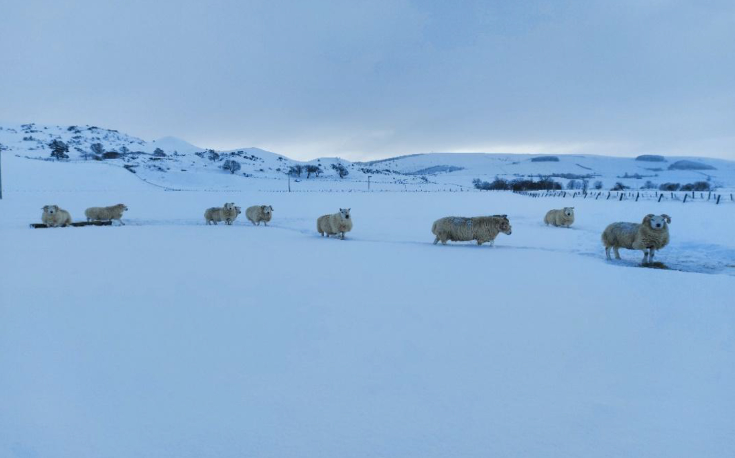
Infection levels appear to be relatively low again this year following a dry and hot summer. Consider testing before treating. Of the twenty positive fluke serology submissions twelve were bovine and eight were ovine. Of the fluke coproantigen ELISA submissions five were bovine, sixteen ovine, two camelids and one caprine. (Note that the fluke serology test is only suitable for cattle and sheep). It is also worth noting that in addition to the positive results there were a further twenty-seven submissions which gave suspicious results in the fluke coproantigen test. Some studies in the literature show that the sensitivity of the coproantigen ELISA is improved by lowering the cut-off without an apparent loss in specificity. Different authors have varied in the lower cut-off that they have suggested for positives, particularly for cattle. We know that the test sensitivity can be adversely affected by pooling so if a result, particularly if it is for a pooled sample, is between 2 and 8% it is possible that infected animals are present.

Geographically, there was a spread of positive liver fluke results from Aberdeenshire in the North to Cornwall in the South.

Fluke serology and coproantigen

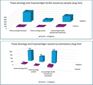
Fluke serology and coproantigen charts by submission and individual samples

New farm team member

We were delighted to welcome Tim Geraghty as an additional recruit to the farm vet team in October. Tim is a European specialist in cattle health and production and has been involved in research and published on a wide range of subjects including fertility, infectious disease and nutrition. He will be working at Axiom on Monday and Wednesday afternoons.
If you wish to speak to a particular vet about one of your cases you can consult the table below to see when our farm vets are working. A duty farm veterinary advisor is always available to speak to on a Saturday morning 9am-12pm:

Vet duty table

Molecular mastitis test

A new rapid, accurate and affordable molecular mastitis test is now available at Axiom. The test detects the presence of six key mastitis pathogens, and the turnaround time is no longer than the following working day. This can allow a targeted course of treatment based upon the pathogens present, aiding responsible use of antibiotics and resulting in less discarded milk. As well as potentially saving money there are benefits for herd health in the long term. The agents detected are E. coli, Staphylococcus aureus, Streptococcus uberis, Streptococcus dysgalactiae, Streptococcus agalactiae and Klebsiella pneumoniae. The cost of the test is £45 & VAT (test code FAMAS).

Animal Health & Welfare Pathway

Axiom is an approved laboratory for testing samples for the parasitology element part of the AHWP for sheep. We don’t supply sampling kits for this purpose, but appropriate consumables can be ordered from us in the usual way.
We can also carry out any of the follow up endemic disease testing for both cattle and sheep. The diseases and conditions to be sampled for sheep include: Border disease (BD), caseous lymphadenitis (CLA), enzootic abortion of ewes (EAE), Johne’s disease, Maedi Visna (MV), toxoplasmosis, tick-borne fever, pulpy kidney, lamb dysentery, ewe nutrition status, lamb nutrition status & trace elements.
For cattle, a biosecurity assessment relating to BVD virus needs to be done in discussion with the farmer. This needs to cover whether it is appropriate for the herd to join a BVD accreditation programme. Membership is not compulsory, as many commercial herds may not be able to meet the rule requirements, particularly the requirement for a minimum three-metre biosecurity gap. Vaccination also needs to be discussed – again it is not compulsory for it to be put in place though it is a good insurance policy until the country is further down the route of eradicating BVD virus. From the BVD check test results coming through the lab most herds appear to be free of infection so could be becoming naïve with time if they are not vaccinating. The impact of a BVD incursion, without the protection from BVD vaccination, could have a serious financial impact on a herd. Although a herd may be closed and appear to be isolated, we have seen breakdowns occurring due to the suspected transfer of virus on equipment or clothing.

BVD Cymru

We are an accredited lab for the Welsh BVD eradication programme. BVD antibody and antigen results will be uploaded if samples are submitted on a BVD Cymru form. As was the case with BVD Free England, there is a small charge for the uploading of the results of 50p per sample for BVD antibody testing and 25p for a BVD antigen test. Please note that all fields on the BVD Cymru submission form must be completed (including the keeper’s phone number and email address) otherwise there is a block on the results uploading.

Discounted Johne’s disease & Neospora testing

Our Johne’s & Neospora Monitoring programmes give farmers access to discounted test rates for whole herd or regular batch testing. There is no membership fee and no set rules to follow. Johne’s serology is from £5 per sample and Neospora serology costs from £6.75 per sample. It works out cheaper than testing through a CHECS cattle health scheme so is ideal for herds that are testing for disease control and management purposes. Advice is provided in the lab report and farmers can be e-mailed a copy if required. Our turnaround times are very fast– often same day but within three working days for both tests. Batch testing herds also get their results in a cumulative spreadsheet. A reminder to test email is sent out for herds on annual testing. Contact us for more information at dsfarm@axiomvetlab.co.uk or on 01626 357776.

Johne’s disease Average Test Values (ATVs)

For herds that are testing using bloods rather than milks for Johne’s serology an average test value can still be calculated but it is not directly comparable to the target used for milks. The Johne’s Action Group has advised that the ATV for bloods should be provided on the declaration form for the herd but state that it has been derived from blood testing. Dairy farmers using Johne’s blood serology can monitor the trend of their herd’s ATVs over time. We will automatically provide an ATV for herds that are testing through the Axiom Johne’s Monitoring Programme (plus any historic values since they started testing through the programme). If one is required when doing a 60 cow screen, please request this on the submission form.

Please use field kits with herd screens

When sending submissions containing 50+ bloods, please ensure that you use a Field Kit containing a polystyrene sample rack/box for the orderly transportation of your samples, remembering to populate the rack in the same order as your accompanying animal ID list. Receiving large quantities of blood samples in a plastic bag or cardboard box is not appropriate or conducive to the efficient handling of such submissions and invariably leads to significant delays in preparation and turnaround times.
Field Kits (filled with the required serum gel tubes) are readily available to order via the following link… https://milab.store.unleashedsoftware.com/

As part of our continuing effort to help improve our services to clients within the Laboratory Division we have put together a short survey which will be sitting live permanently for anyone to complete whenever they have a few minutes spare. This can be filled in by anyone who uses any of our services and we encourage feedback to help us understand what we are doing well and where we need to make improvements. The QR code and link to the survey will remain live as a continuous tool to enable us to always gather feedback. If anyone has any questions about this they are welcome to contact either of the Quality Managers at the Laboratories, Claire Richardson for Axiom Veterinary Laboratories and Susan Reeve for Finn Pathologists. Thank you in advance for helping us to improve our services.

https://www.surveymonkey.com/r/Laboratory_Satisfaction_Survey

Interesting cases in October

Cattle
Abortions

  • Bacillus licheniformis was cultured from stomach contents of aborted bovine foetuses this month. Bacillus licheniformis is a known cause of abortion in cattle, which may be associated with ingestion of poor quality silage, silage effluent, contaminated feed, trough water and bedding.
  • There were five cases of bovine abortion attributed to Salmonella infection, all of which were serotyped as Salmonella Dublin, a host adapted strain of Salmonella in cattle.
  • There was one bovine abortion case where foetal fluid tested positive for antibodies to Neospora caninum indicating the calf had seroconverted in utero. The finding however is not sufficient to diagnose neosporosis as the cause of the abortion – that requires histopathology of foetal heart and brain.
  • Samples were submitted from two aborted dairy calves from the same holding. The veterinary practitioner had noted that there was subcapsular haemorrhage of the foetal kidneys. Both foetuses were positive for Q fever (Coxiella burnetii) by PCR. In this case one of the two foetuses had a low positive result for BVD antigen in foetal fluids. Positive antigen results on foetal fluid may be due to an in utero BVD virus infection (e.g. following a transient BVD viraemia), the use of a live BVD vaccine in a pregnant animal or it could be a false positive result. Further investigation of the herd’s BVD status was recommended e.g. bulk milk BVD serology and BVD virus PCR test plus antibody check tests on young stock groups.
  • Corynebacterium provencense was isolated from a sample submitted for investigation of mastitis. Corynebacterium spp. are frequently detected in milk samples, but it is uncertain whether their presence is due to infection, non-pathogenic colonization or contamination. Corynebacterium spp. have been detected in case of subclinical mastitis and clinical mastitis but may also be isolated from the milk of healthy cows with no subclinical or clinical signs of mastitis.

Respiratory disease

  • Postmortem tissues were received from a six-month-old beef calf with a history of respiratory signs, and which was the third of the group to die. There had been a variable response to treatment with oxytetracycline and meloxicam in others in the group showing signs of pneumonia. There was marked consolidation of both cranioventral and caudal lung. Small numbers of lungworm were observed in the airways. Pasteurella multocida was isolated in cultures of lung swabs and the isolate demonstrated resistance to multiple antimicrobials in vitro. It was considered that the isolate was likely to be significant in the case of pneumonia and that P.multocida often acts as a secondary agent. Histopathology identified severe, chronic and ongoing suppurative broncho interstitial pneumonia. There was evidence of extensive airway damage, an adult lungworm was seen in one section, and there was a suppurative bronchopneumonia consistent with the Pasteurella multocida infection. It was noted that the observed pathology could all be attributed to lungworm with secondary bacterial infection. However, it was not possible to completely rule out a previous or concurrent viral insult (e.g. BHV 1, RSV and PI3), inhaled irritant or toxin in this case.
  • Mannheimia haemolytica was thought to the likely cause of the gross and histopathological changes in the lung tissue of an adult cow which died following a short course of illness and scour suspected to be due to salmonellosis.
  • A Holstein cow which was recovering from a case of mastitis presented with condition loss and a bilateral ocular discharge. IBR virus was detected by PCR testing of a nasopharyngeal swab.
  • Parasitic pneumonia in cattle due to D. viviparus was frequently diagnosed in October. In one herd of 220 dairy cows which had been housed three weeks previously there was very frequent coughing. Cows were otherwise reported to be well in themselves. There had been no recent anthelmintic treatments given. Baermann’s examination of a pooled faecal sample from three of the cows was positive for the presence of larvae resembling D.viviparus.
  • Postmortem tissues were received from an adult dairy cow from a farm where there was a history of adult milkers dying with severe respiratory signs. Lung histopathology revealed severe, chronic and ongoing changes with frequent nematode larvae seen. The pathology was considered consistent with ongoing lungworm infection, where adult worms have been eliminated (no adult worms were seen in the examined section). Acute, fatal exacerbations of disease can occur during the period of recovery.
  • Two Guernsey calves at pasture which were losing condition and coughing and had excessive upper respiratory tract noise were positive for antibodies to D.viviparus on serology indicating they had been exposed to lungworm.
  • Salmonellosis was the likely cause of death in a dairy heifer calf of less than two weeks of age. It was one of several young calves to die recently on the same farm. The main finding at postmortem was lung pathology suggestive of bronchopneumonia and it was noted that the liver had a yellow tinge. Multiplex PCR testing for respiratory pathogens did not detect any of the common respiratory viruses or bacteria. Histopathology revealed a moderate, suppurative bronchopneumonia with intralesional bacteria which stained as gram-negative. Intravascular fibrin thrombi were seen in liver tissue and there was evidence of a suppurative serositis in the intestinal sections, although autolysis prevented full evaluation of gut tissues. Selective culture of lung tissue isolated a Salmonella sp. which was identified by serotyping as Salmonella Typhimurium.

Gastrointestinal disease

  • E.coli F5 (K99) antigen was detected in faeces of a four-day-old Aberdeen angus cross calf. It was the fourth calf to die on the farm at less than five days of age after acute signs of scour.
  • Coccidiosis was diagnosed in a yearling Belted Galloway animal which had shown signs of scour and loss of condition in the previous five days with strands of rectal mucosa observed in the faeces. There was a coccidial oocyst count of 2000opg of which 100% were of the pathogenic Eimeria species, Eimeria bovis.

Skin and eye disease

  • A vulval mass from a heifer removed by excisional biopsy was identified as a likely fibropapilloma by histopathology.
  • Fusobacterium necrophorum (foul in the foot) was suspected to be involved in an outbreak of severe foot lesions involving sloughing of the hoof wall in three adult cows on the same farm. Biopsy and histopathology identified evidence of necrosis of the laminae with accompanying bacterial infection and inflammation. The features are considered typical of a Fusobacterium necrophorum infection. No Treponemes spp. were detected by PCR testing in this case.
  • Moraxella bovoculi was isolated in a case of Infectious Keratoconjunctivitis in dairy cattle which were showing signs of melting corneal ulcers. Although this bacterium is associated with cases of IKC, it can also be found in the eyes of healthy cattle.

Systemic and miscellaneous disease

  • Babesiosis was diagnosed by identification of blood parasites in a blood smear from a four-year-old stabiliser cow which was down and had evidence of haemoglobinuria. Babesiosis was reported by the vet as being previously unknown in this area (North-East England).
  • OvHV2, indicative of MCF in cattle, was detected by PCR testing in at least four cases this month. One case was in a Belted Galloway bull which had showed signs of corneal oedema for two weeks which progressed to pyrexia , anorexia , lethargy and balanoposthitis.
  • Very high CK levels (>20,000UL, RI 0-200U/L) and AST levels (1742U/L RI 0-100U/L) indicative of severe myositis most likely due to trauma were measured in a yearling heifer which had become stuck in a ring feeder and was unable to rise after being freed.


Sheep and goats

Gastrointestinal disease

  • Johne’s disease was diagnosed by serology in a two-year-old ewe which presented with unexplained weight loss. On another farm a group of twelve thin ewes were sampled for investigation. Both pooled faecal samples tested positive for MAP (Mycobacterium avium paratuberculosis) by PCR indicating that Johne’s disease was present in the group. One of the pooled samples had a suspicious result in the fluke coproantigen ELISA indicating that liver fluke infestation may also be contributing to the condition.
  • Yersinia enterocolitica was isolated from faecal samples from five-month-old Welsh Mountain lambs which were suffering from scour and an increased incidence of sudden deaths. Faecal worm egg counts had been repeated on several occasions with negative results. Yersinia enterocolitica is an uncommon cause of enteritis in sheep. Wild birds and rodents are thought to be potential sources of infection. As yersiniosis can occasionally be associated with underlying concurrent disease or deficiency, trace element screening to investigate possible nutritional deficiency was suggested.
  • Eggs of the lancet fluke (Dicrocoelium dendriticum) were detected in faecal samples from sheep in the North of England. Although considered a rare finding, lancet fluke is recognised more commonly in some localised parts of the UK (Cornwall, Inner Hebrides). The parasite has a complex life cycle which involves two intermediate hosts: a snail and an ant. Infections in sheep are most commonly subclinical but heavy burdens can lead to weight loss, anaemia, bile duct fibrosis and liver cirrhosis. They are most often identified as a (usually) incidental finding at postmortem or as in this case eggs can be identified in faeces using a flotation method. (D.dentriticum eggs are much smaller than either rumen or liver fluke eggs)
  • A pooled faecal sample tested four weeks following triclabendazole treatment was positive in the fluke coproantigen ELISA indicating treatment failure and suspected triclabendazole resistance.

Skin and eye disease

  • A pygmy goat which presented with a large fluid filled submandibular swelling tested positive for Caseous lymphadenitis (CLA) on serology. Corynebacterium pseudotuberculosis was isolated in culture of swabs from two rams from the same flock with abscesses.
  • Streptococcus dysgalactiae was cultured from vulval swabs from ewes showing signs of ulcerative vulvitis. Three ewes were affected in a small hobby flock of fifteen. The ram which was introduced six weeks previously had no visible signs of ulcerative posthitis. It was noted that animals on a high protein diet such as lush pasture can be predisposed to this condition. Ewes are usually less commonly and less severely affected with ulcerative vulvitis than male sheep are with posthitis.

Systemic and miscellaneous disease

  • Around seven weaned lambs from a group of 160 died and signs of septicaemia and pneumonia with excess fluid present in the pleural cavity were seen at gross postmortem examination. Histopathology of lung identified moderate, multifocal, acute, necrotising interstitial pneumonia with intralesional bacterial colonies. This appearance is characteristic of septicaemic pasteurellosis (in this case likely Bibersteinia trehalosi). Associated stressors include weaning, handling, transport, mixing/moving and weather-related factors. There were at least seven other cases of systemic pasteurellosis in sheep this month all with a history of sudden deaths and in one case recent long- distance transportation.

Clusters of bacteria

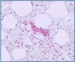
Clusters of bacteria (Biberstenia trehalosi) with admixed degenerate leukocytes in the lung of a sheep with systemic pasteurellosis (HE)

Sheep systemic pasteurellosis

Sheep systemic pasteurellosis: Bacteria are gram negative (pink coccibacilli) on Gram stain.

  • Blood samples were received from a group of ewes which were described as ‘varying degrees of thin’ despite having access to good quality grazing. Four of the five ewes blood tested were positive for antibodies to Maedi-visna and this was considered the likely cause of the poor condition.

Camelids

  • A high number of strongyle eggs (8800epg) were observed in a dung sample from one adult male alpaca in a group of three. A second animal in the group had a strongyle count of 350epg and the third had a negative strongyle egg count. Haemonchus identification was carried out on the sample with the high count and 97% of the observed oocysts were identified as Haemonchus sp. This highlights the very variable worm egg counts that can occur in groups of livestock with haemochosis.
  • Staphylococcus aureus was isolated from a skin swab from alpaca showing signs suspicious of pyoderma with papules, raised nodules and evidence of ulceration on chest and groin. The animal had a previous history of dermatitis due to mite infestation.

Pigs

  • On-farm postmortem of a grower pig identified extensive fibrinous percarditis, pleurisy and peritonitis. Pasteurella multocida was isolated in cultures of lung tissue. As in cattle and sheep P.multocida is often present in the upper respiratory tract of healthy pigs. However, it can cause an exudative bronchopneumonia, sometimes with pericarditis and pleuritis, often secondary to other underlying disease. In this case PRRS virus was detected in the lung tissue by PCR testing. PRRS infection is a known risk factor for increased mortality due to respiratory disease in weaner and growing pigs.
  • Rotavirus was detected by PAGE testing of a faecal sample from four-week-old diarrhoeic piglets from a smallholding of ten sows where one litter had signs of scour. Older piglets were bright and unaffected. The finding of Rotavirus was considered likely to be of significance in this case.
  • Staphylococcus hyicus, the cause of greasy pig disease was isolated from lesions on fattening pigs presenting with severe ear tip necrosis. These bacteria can penetrate through small abrasions or lesions on the ear and proliferate in damaged tissue.

Deficiencies and toxicities

  • There were at least 40 cases of suspected acorn toxicity in cattle in October and at least twelve in sheep. Most of the submissions had a history of clinical signs suggestive of toxicity with possibility of access to acorns and /or acorns found in the rumen at postmortem examination. Biochemistry typically identified very high urea and creatinine levels consistent with renal dysfunction/renal failure.

Kidney from a sheep with acorn toxicity.

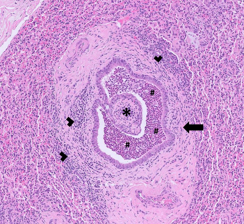
Kidney from a sheep with acorn toxicity. Tubules are often dilated and filled with eosinophilic fluid, haemorrhage, necrotic debris and degenerate neutrophils (arrows). There is also multifocal interstitial information (circle)

  • An eight-year-old Holstein cow showed signs of a gradual decline in milk yield followed by inappetence and dullness. Neurological signs including exaggerated blinking, twitching of the head, ataxia and hypermetria were observed. No ketones were identified on dipstick testing of the urine. Testing indicated the animal was hypomagnesaemic with a magnesium level in serum of 0.62 mmol/l (0.70-1.3mmol/l) and this was thought to account for the clinical signs in this case.
  • Copper toxicity was diagnosed postmortem in a five-month-old lamb and an eight-month-old lamb on separate submissions. Kidney copper levels were well above normal levels at 2580umol/kg DM and 4690umol/kg DM respectively (RI 0.00-787umol/kg DM) consistent with copper toxicity in both cases.
  • Evidence of Zinc deficiency was found in blood samples from six llamas tested to investigate trace element status. Some animals in the herd were of lower-than-expected body condition, despite actively monitoring parasite status and several individuals had poor skin condition with scurfy skin and some chronic skin changes.

Other Species

  • Several piglets from a litter died at 24 to 48 hours of age, following a period of inappetence and lethargy. The dam was also unwell. There had been some response and some recoveries after administration of intramuscular oxytetracycline. PME of one piglet that died a few days after the others but with the same clinical signs detected splenic enlargement, jaundice and purple discolouration of the skin. Erysipelothrix rhusiopathiae was isolated in moderate pure growth from a liver swab. Erysipelas is rare in such young piglets but it has nonetheless been reported in this age group and was considered likely to have been significant.
  • Pericarditis and perihepatitis were seen on PME of five to six-week-old rare breed poultry chicks and the likely cause was E. coli infection after its isolation in moderate, pure growth from a thoracic swab.

Follow us on Facebook

https://www.facebook.com/Axiomfarmvetlab/

This newsletter is sent by e-mail to each vet practice but if you would like a copy sent to your individual e-mail account please contact us at dsfarm@axiomvetlab.co.uk and we can add you to our circulation list.

Related Posts