Email Us
admin@axiomvetlab.co.uk

admin@axiomvetlab.co.uk
(01626) 355655

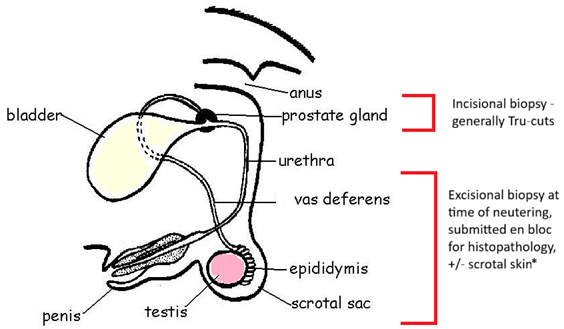

Image adapted from: https://commons.wikimedia.org/wiki/File:Male_repro_system_labelled.jpg
*It’s not necessary to send scrotal skin unless you have a concern about a scrotal lesion. However, if you are interested in scrotal lesions, particularly potential neoplasms, you could you consider inking the margins (see Finn fact sheet 102).
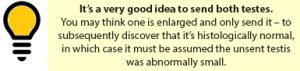
Testes/epididymis and spermatic cord/pampiniform plexus
Prostate gland
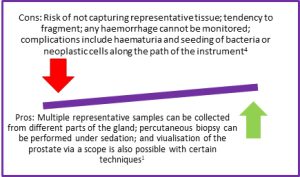
A few diagnostic methods are used to assess canine prostatic gland, but biopsy is the gold standard and achieves a diagnosis in approximately two thirds of cases3. Sampling may be surgical or percutaneous1,4. Biopsies may be Tru-cuts or wedge resections and there are some advantages and disadvantages to be aware of, outlined in Fig 1. Main contraindications to prostatic biopsy are acute prostatitis and potential prostatic abscess1.
Fig 1. Pros and Cons of Tru-cut prostatic biopsies
This section includes examples of the more common lesions we see.
Fig 2: Testicular seminoma. This presents as solid sheets of atypical cells reminiscent of a round cell tumour. Inset: Higher power highlights atypical germ cells with one atypical mitotic figure (yellow arrow). Grossly, these tumours are often off-white, soft and slightly bulging on cut section.
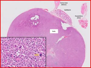
Fig 3. Sertoli cell tumour. Composed of supportive cells from the seminiferous tubules. Approximately 20-30% of affected dogs manifest signs of feminization due to hyperoestrogenism. The majority of Sertoli cell tumours are benign – metastasis is more likely to occur with larger tumours, and involves the sublumbar and pelvic lymph nodes, with potential dissemination to various internal organs.
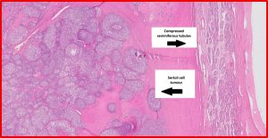
Fig 4. Sertoli cell tumour. The arrangement is more tubular, or at least distinct nesting, rather than sheets, with palisading on the basement membrane (yellow arrow). The cells are also spindle shaped or elongated rather than round. Grossly, these tumours are white and firmer than seminomas due to more abundant stroma.
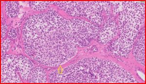
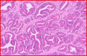
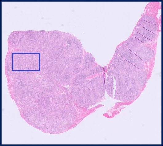
Fig 5. Prostate gland. This is histologically normal but, as the prostate gland was visibly enlarged, the changes are consistent with benign prostatic hyperplasia.
Mammary Glands I. Indications for biopsy/pathological evaluation Mammary lesions are among the most common samples…
Organ of the month: Oral I. Indications for for oral biopsy Oral biopsies are among…
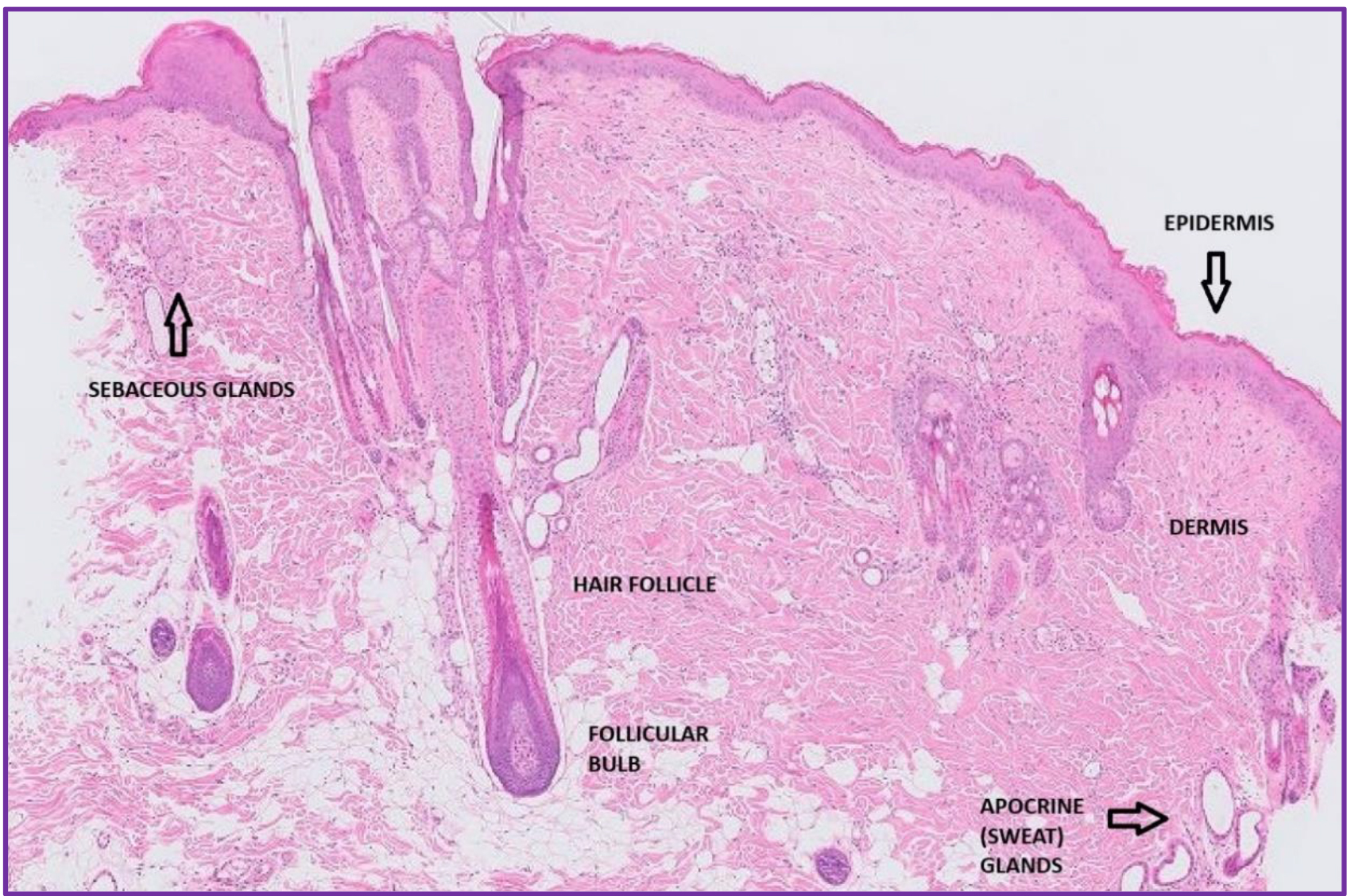
Organ of the month: Skin I. Indications for skin biopsy Skin biopsies account for a…
PART I – The Female Reproductive Tract I. Indications for biopsy/pathological evaluation As a reminder,…
The great explorer of the truth, the master-builder of human happiness no one rejects dislikes avoids pleasure itself because it is pleasure but because know who do not those how to pursue pleasures rationally encounter consequences that are extremely painful desires to obtain.
Read More


