Farm October Newsletter 2026

Dedicated farm line – 01626 357776

Follow us on Facebook

https://www.facebook.com/Axiomfarmvetlab/

This newsletter is sent by e-mail to each vet practice but if you would like a copy sent to your individual e-mail account please contact us at dsfarm@axiomvetlab.co.uk and we can add you to our circulation list.

Would you like to send the Axiom farm vets photos of PMEs to allow us to assist with sample selection and diagnosis? Please send photos by WhatsApp to 07944 649 222.

Note that we may wish to include photos sent to this number for the interesting cases section of this newsletter. If you do not wish your photo to be used, please let us know at the time of sending.

Email us with your additional test requests

You may find this is a more efficient way of making requests than phoning the farm team, saving you time in your busy day. Our farm team also find it a more efficient way of dealing with your requests.
The email address for test requests is: dsfarm@axiomvetlab.co.uk 

Shamonda virus

There have been outbreaks of infection involving new variants of Shamonda virus in Switzerland, Germany, France and more recently Jersey this year. There is a risk that it will cross the Channel. It is very closely related to Schmallenberg virus, is also transmitted by Culicoides midges and causes very similar signs of milk drop, diarrhoea, sometimes pyrexia, reduced conception rates, abortions and congenital defects may also occur depending on the stage of pregnancy infection occurs at. It is possible that the viraemic phase is short lived, as with SBV, which is approximately two to six days, but blood samples taken during that period can be tested by PCR for the virus. Abortion material can also be tested (e.g. brain, spleen) and we are hoping to have a PCR test in place very soon. Serology is not currently possible. Previously exposed animals are likely to test as antibody positive in the SBV ELISA.

Bluetongue testing guidance

Please note that, due to SAPO regulations, we cannot accept samples from animals showing signs suspicious of Bluetongue infection, being tested for Bluetongue or that have recently tested positive for Bluetongue. If such samples are received, they will have to be destroyed. We can accept samples from herds or flocks which are currently undergoing testing for BTV or are showing signs consistent with BTV infection provided there is no suspicion of bluetongue virus infection in the individual being tested. We are happy to discuss cases over the phone before submission of samples. Clinical suspicion of BTV infection must be reported to APHA.  For the latest information and updates on BTV in the UK please visit the following link:

Bluetongue: latest situation – GOV.UK
Bluetongue in dogs

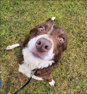
There is a general lack of awareness that dogs can become infected with bluetongue virus and unfortunately in a small number of cases it can have severe consequences. Most cases involve subclinical infection but pregnant bitches are at highest risk, particularly from mid pregnancy onwards. They can abort and tend to die shortly afterwards due to severe pulmonary oedema. Bleeding disorders, resulting in spontaneous haemorrhages, can also occur occasionally in dogs due to infection. The main source of virus for dogs is believed to be from eating infected raw meat. It is therefore advisable to avoid feeding pregnant dogs raw meat whilst there is bluetongue virus in the country. Farmers should take steps to prevent farm dogs from being able to access ruminant carcases, abortion material and placentas. Although there is some evidence that midges may occasionally be the source of the virus for dogs they much prefer biting cattle and sheep.
We suspect there have been some bluetongue cases in dogs in Wales. One involved two bitches on the same premises, which aborted in mid pregnancy and then died and there had been evidence of severe pulmonary oedema on radiographs. They had eaten part of a sheep that was believed to have died from bluetongue. There was also another possible case, also in Wales, involving multiple dogs, where one died acutely following a coagulopathy, others were ill and the dogs involved had been fed carcase meat. Whilst bluetongue is a notifiable disease in livestock it is not in dogs so unfortunately it has not been possible to confirm the diagnosis in these cases as testing is being restricted to ruminants and camelids.

Axiom Farm Vets

If you wish to speak to a particular farm vet about one of your cases, you can consult the table below to see when our vets are usually available. A duty farm veterinary advisor is always around on a Saturday morning from 9am-12pm.

Farm Histopathology

We offer farm histopathology on postmortem samples, and on biopsy samples from live animals under the code FPM01. Up to three different tissues can be processed under this code and extra tissues can be processed on request. Where different lesions/stages of disease are seen in one organ (e.g. pneumonic lungs) multiple small sections can be submitted as one tissue type and this will not incur additional costs. For best results only perform histopathology on tissues from fresh carcases and ensure samples are transferred into fixative as soon as possible after sampling. It may not be possible to process samples which have been submitted as fresh tissues if they appear severely autolysed on arrival. Formalin containing histopathology pots can be ordered with other consumables through the website. Scan the QR code to access our ‘How to get the most out of your large animal histopathology service’ factsheet. Further tips on sampling for histopathology can be found in the ‘Histopathology: points to consider’ and ‘Sampling for respiratory disease’ factsheets.

Axiom Farm Factsheets

Animal Health and Welfare Pathway Parasitology

Axiom is an approved laboratory for testing samples for the parasitology element part of the Animal Health and Welfare Pathway (AHWP) for sheep. For both the pre-treatment and post-treatment elements of the drench efficacy check we require a minimum of ten samples from the group of sheep being tested and no more than fifteen to make up the composite sample. In some case, for example very small flocks, there may be insufficient animals in the group to sample. Please consult the AHWP scheme guidance for further information for exceptions to the sampling rules and let us know if these exceptions apply when submitting the samples.

As with any faecal worm egg counts, results for AHWP submissions must be interpreted along with the clinical presentation. Many factors including age, nutritional status, previous exposure/immunity, species of parasite and faecal consistency will affect the results obtained.

We have a specific submission form which you can use for AHWP parasitology and you can find this on our website at:

AXIOM VET LABS LTD SHEEP WORMING TREATMENT CHECK SUBMISSION FORM Issue 2. 01.04.2025.indd

Please fill in as many details as you can including treatment and sampling dates and any clinical signs where relevant.

To allow us to provide WEC counts of sufficient sensitivity for the AHWP drench efficacy test we use OvacyteTM digital technology. We participate in regular QA (Quality Assurance) testing of both McMaster and OvacyteTM. Typical variation in QA testing is extremely high across all labs taking part (1 SD (Standard Deviation) typically around 45% of the mean) and so it is anticipated that comparison results between practices and labs will vary widely. This could be due to very uneven distribution of eggs within a faecal sample, which could be exacerbated by pooling of samples from animals with very differing counts. Interpretation of counts, including drench checks, should be made with this level of variation in mind, particularly in results close to cut-off points. There could be cases where, based on clinical presentation in a group, the appropriate decision might be to re-test or to treat the group even if the count is only moderate (given variation can be so high). These cases might not be suitable for ‘pathway’ drench checks however (or at least care would be needed in interpretation). Whilst a 90% reduction is the recommended cut-off for ‘effective treatment’, samples with a low or moderate pre- treatment count may not achieve this despite an effective treatment. A high count in the post-treatment sample, assuming the timing, collection and submission method has been following appropriately, would always indicate treatment failure.

Further information on parasite control in sheep and how to perform drench efficacy checks is available from www.scops.org.uk

Feedback on our services

As part of our continuing effort to help improve our services to clients within the Laboratory Division we have put together a short survey which will be sitting live permanently for anyone to complete whenever they have a few minutes spare. This can be filled in by anyone who uses any of our services and we encourage feedback to
help us understand what we are doing well and where we need to make improvements. The QR code and link to the survey will remain live as a continuous tool to enable us to always gather feedback. If anyone has any questions about this, they are welcome to contact either of the Quality Managers at the Laboratories, Claire
Richardson for Axiom Veterinary Laboratories and Susan Reeve for Finn Pathologists. Thank you in advance for helping us to improve our services.

https://www.surveymonkey.com/r/Laboratory_Satisfaction_Survey

Interesting cases for September

Cattle
Abortions/mastitis

  • There were at least three bovine abortion cases this month where foetal fluids tested positive for BVD antigen in the ELISA on foetal fluids (fluids within thoracic /abdominal cavity). In all these cases, abortion occurred in late pregnancy. There was one foetus in which the fluid also tested positive for antibodies to Neospora caninum in the Indirect Fluorescent Antibody Test (IFAT), indicating both BVD viraemia and seroconversion to Neospora in-utero in that foetus.
  • There were several cases in which foetal tissues tested positive for Neospora caninum by PCR testing. The preferred samples for this test are foetal brain or placenta (with cotyledon).
  • One farm was experiencing an abortion storm with twelve cows aborting over three weeks in late gestation. Ten aborted cows were tested for antibodies to Neospora caninum and all were positive, raising the suspicion of point source (exogenous) neosporosis.
  • Coxiella burnetii was detected by pooled PCR testing of a vaginal swab and placental cotyledon from an aborted cow using the abortion PCR package screen. Detection of Coxiella burnetii may be an incidental finding and additional evidence of foetal infection, for example demonstration of placentitis by histopathology, is required to make a diagnosis of abortion due to Q fever.
  • There were several cases of abortion due to Salmonella Dublin this month. One affected dam passed black tarry dung before aborting at two months gestation.
  • Staphylococcus aureus was implicated in one case of bovine abortion in the seventh month of gestation. Cultures of the placenta yielded profuse growth of Staphylococcus aureus raising the possibility of aureus associated placentitis. Staphylococcus aureus is recognised as a sporadic cause of abortion in cattle and is most likely to be due to haematogenous spread following infection in the dam.
  • There were several cases of mastitis due to Trueperella pyogenes diagnosed in September. Trueperella (formerly Arcanobacterium) pyogenes can cause an acute purulent form of mastitis. Infections occur most frequently in dry cows or heifers before calving. Infections may occur in lactating cows after a teat injury. pyogenes is commonly involved in the aetiology of ‘summer mastitis’.
  • Pasteurella multocida was isolated from one of a number of mastitic milk samples from a farm with persistent high cell counts and mastitis Pasteurella species infections are a rare cause of clinical mastitis. The organisms are usually commensals in the oral cavity and upper respiratory tract. Haematogenous or spread via the lymphatics may be the route of spread to the udder under certain conditions. Suckling calves may also be a source of infection. Cow to cow spread is suspected to occur so it is advised that affected cows are removed from the herd as there is usually a poor response to treatment. Cows can become systemically ill and die due to the production of endotoxins by the organisms. Affected cows should be milked last or with a separate cluster until they can be removed from the herd. Cows with teat injuries appear to be at higher risk of infection so maintenance of good teat condition is advised in the prevention.
  •  

Respiratory disease

  • IBR virus was detected by PCR testing of nasopharyngeal swabs from an adult Holstein-Friesian cow which was recumbent, open-mouth breathing and drooling. In another case of IBR in adult dairy cattle the virus was detected by PCR of two separate pools of nasopharyngeal swabs, one from recently bought-in cattle and another from homebred animals. Respiratory signs, milk drop and scour had been observed and two abortions had occurred in affected animals. There were at least four other cases of IBR diagnosed by PCR testing in September, one was on a beef unit, and four animals there had died with severe respiratory illness. In that case IBR virus was detected by PCR of pooled lung and tracheal tissue sampled during on farm postmortem.
  • On one dairy unit, the farmer had observed that young calves appeared to go through a period of nasal discharge and coughing. A nasopharyngeal swab was taken from an acute, typical case and PI3 virus was detected by PCR testing indicating evidence of active virus Although PI3 is recognised as a cause of respiratory disease in young cattle, some animals may show few or no clinical signs.
  • RSV was detected in a pool of three nasopharyngeal swabs from twelve-week-old calves with clinical signs of pneumonia, indicating active RSV infection. RSV is probably the most significant viral cause of respiratory disease in young cattle.
  • A three-day-old dairy heifer calf showed signs of respiratory distress and died a short time after. The owner was concerned that there may have been incorrect placement of a stomach feeding tube. On histopathology of lung tissue bronchioles were observed to contain a mix of mucoid material, neutrophils, pigmented material, squames and some macrophages, with some fluid like material also present consistent with meconium aspiration and bronchopneumonia. Whilst aspiration of meconium in calves is not believed to have primary clinical significance, in this case there were significant quantities of material with a neutrophilic response suggesting clinical significance and associated bronchopneumonia. The small amounts of fluid material may have been indicative of aspiration of milk, but overall, this was considered a minor feature and may have been secondary to respiratory embarrassment associated with the pneumonia. If it was assumed that the findings were representative of all affected calves, then it was suggested that attention should be addressed to managing dystocia/difficult calvings.
  • A Holstein- Friesian dairy cow was one of several in the herd which showed respiratory signs and mild drop. This cow also developed a swelling in one of the hock joints. Initial testing for antibodies to Mycoplasma bovis during the acute phase gave a high negative result of 57% (>/=60% is a positive result). Resampling after three weeks found a high positive result of 214% in the Mycoplasma bovis ELISA indicating seroconversion consistent with recent exposure. This was a flying herd and it was considered that bovis may have been introduced recently. Further testing to identify underlying factors which could contribute to disease due to Mycoplasma bovis in the adult herd, such as energy, protein or Vitamin A deficiencies, was discussed with the submitting vet.
  • Fixed lung tissue was received from a month-old-calf which had been treated multiple times for pneumonia and scour but had failed to respond and was euthanased for Consolidation of the right cranial lung tissue and purulent material within the airways was observed. Histopathology identified necrosuppurative, bronchointerstitial pneumonia with features strongly suggestive of a Mycoplasma bovis-associated pulmonary insult.
  • Pasteurella multocida was detected on PCR testing of lung tissue from a six-month-old Limousin cross calf, which had shown signs of respiratory distress at grass and had collapsed and died as the farmer attempted to herd it back to the farmyard for A significant amount of fibrin was seen on the pleural space at postmortem examination. The findings were suggestive of a primary pasteurellosis though no obvious stress factors (e.g travel or extreme weather conditions) were identified. A liver sample was tested to assess trace element status, and the selenium level was found to be below the reference interval at 7.33µmol/kg DM (RI = 11-22.6µmol/kg DM). It was considered that selenium deficiency may have been a contributory factor in the case.
  • Trueperella pyogenes was isolated in cultures of swabs from abscesses seen in the lungs of a five-year-old bison which died suddenly at pasture. Ulceration of the soft palate, liver abscessation, collapse of the cranial lung lobes and a small amount of purulent discharge in the airways were observed at postmortem examation. pyogenes is most usually a secondary opportunistic pathogen in lungs, so its isolation did not identify it as a likely cause of sudden death. Histopathology of lung tissue revealed marked diffuse lung oedema with extensive fibrin exudation. While pulmonary congestion and oedema are often considered non-specific changes and may represent agonal phenomena, the presence of fibrinous exudation suggested a peracute pathological process and in conjunction with intralesional bacteria, raised the possibility of a septicaemic event. Examination of further tissues particularly of kidney and brain was recommended but unfortunately these were not available in this case. Evaluation of the brain is especially relevant in cases of sudden death, as significant lesions may be present despite limited findings in other organs and may provide important clues regarding the underlying cause of death. Similarly, renal lesions may help support or refute the possibility of a systemic infectious process.

Gastrointestinal disease

  • A six-month-old Aberdeen Angus cross calf presented with mild pyrexia and blood The coccidial oocyst count in a faecal sample was >50,000opg of which 87% were identified as the pathogenic species Eimeria zuernii, confirming coccidiosis in this calf.
  • A faecal sample were received from animals on a calf rearing unit in which calves of one to two weeks of age were developing profuse water diarrhoea and three had died. The sample tested positive for Cryptosporidium antigen and Salmonella Coeln was isolated in cultures. S. Coeln is a rarely identified serotype in cattle, which has been associated with contaminated feedstuffs.
  • Salmonella Dublin was isolated in multiple cases of gastrointestinal disease in cattle in September including in an eleven-day-old calf with pyrexia, severe scour and dehydration and in a four-year-old Charolais bull which had shown signs of diarrhoea and weight loss in the previous two weeks.
  • coli F5 (K99) antigen was detected in at least three cases of diarrhoea in neonatal calves.
  • Liver fluke eggs were seen on microscopic examination of two samples from yearling cattle which were not thriving.
  • Johne’s disease was diagnosed on serology testing of a twelve-year-old Limousin bull which was losing condition, in an eighteen-month-old Holstein cross bullock which had weight loss, watery diarrhoea and bottle jaw and in a fifteen-month-old Limousin cross bullock with signs of ill-thrift and scour. There were many more cases in the typical age range of three to five years old.
  • A two-year old British Blue cross cow showed signs of mild pyrexia and tenesmus with blood and mucous present in the dung. In house parasitology had found no evidence of gastrointestinal parasitism or The animal had a strong positive result for BVD antigen on ELISA, confirming BVD viraemia and likely mucosal disease. It also tested positive for antibodies to Johne’s disease on serology.

Skin and eye disease

  • Cutaneous actinobacillosis was the suspected cause of a skin lump on the flank of a three-year-old Highland cow. The animal had been previously showing signs of a laryngeal problem suspected to be due to actinobacillosis. Histopathology of a section of the skin mass identified a focally extensive, chronic, marked pyogranulomatous and lymphoplasmacytic dermatitis/cellulitis with intralesional bacteria and Splendore-Hoeppli reaction (bright pink-staining eosinophilic material surrounding infectious agents or foreign bodies). The changes on histopathology were considered highly suggestive of Actinobacillus lignieresii infection, which may be confirmed by cultures of fresh tissue, though bacteriology was not carried out in this case. Actinobacillus lignieresii can cause ‘wooden tongue’ in the mouth, as well as cutaneous actinobacillosis in the skin. Trauma usually allows its entry.
  • Moraxella bovoculi was isolated in aerobic cultures of an eye swab from a dairy calf of less than one week of age which was one of several calves affected in the The finding on culture was considered significant and indicative of Infectious Bovine Keratoconjunctivitis (New Forest eye), though this condition is unusual in such young calves. M. bovoculi was isolated in one other submission, in this case the problem was being seen year-round in cattle from weaning age to adults.
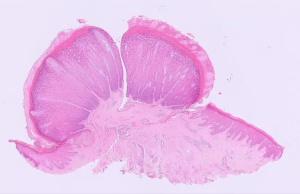
  • Biopsy samples from three Holstein-Friesian dairy cattle from a herd which was experiencing an outbreak of suspected teat papillomas were submitted for The histopathological appearance of all the samples confirmed changes typical of teat papillomas of epithelial hyperplasia, hyperkeratosis, acanthosis, and koilocytes (abnormal squamous epithelial cells) (Figure 1). In most cases teat papillomas are caused by bovine papillomavirus infection (including BPV-6, 7, 9 and 10), and the appearance in all three samples in this case support a viral aetiology.

Figure 1: A papilloma on the teat of a cow showing marked epidermal hyperplasia and hyperkeratosis.

Systemic and miscellaneous disease

  • There were several diagnoses of babesiosis in cattle this month, including cases from Devon and Dorset.
  • BVD viraemia was detected in three animals on pooled BVD PCR testing followed by BVD antigen ELISA testing of individuals from each of the three positive A PI hunt had been initiated in this herd following observation of rising BVD antibody results on bulk milk tank serology. This was one of a number of herds where likely PI animals were identified following pooled PCR testing.
  • Chronic pyelonephritis was identified on histopathological examination of renal tissue from an in-calf adult Limousin cow which had shown signs of abdominal pain. Biochemistry had previously identified significant azotaemia suggestive of renal dysfunction, raised globulins consistent with chronic inflammation and hypoalbuminemia which may have been the result of protein losing nephropathy. Gross pathology of the kidney was consistent with pyelonephritis and this was confirmed by histopathology which found evidence of chronic, ongoing extensive changes in the kidney tissue. Pyelonephritis may be caused by ascending urinary infections or by haematogenous spread. The distribution of the lesions in this case was suggestive that it was the result of previous/ongoing bacteraemia with embolic spread to the kidneys. The original focus of inflammation elsewhere in the body (e.g. mastitis, metritis, foot abscess, tooth abscess etc.) may no longer have been present at the time of death.
  • Trueperella pyogenes was isolated from a joint fluid sample in a pre-weaned Holstein The farm was experiencing an outbreak of joint ill cases, with no evidence of navel infections observed.
  • There were at least four cases of malignant catarrhal fever recorded in September, all of which had some typical clinical signs such as pyrexia, corneal opacity and crusting of the nasal planum and one included a history of co-grazing with sheep. Close contact with sheep is the main risk factor for MCF as sheep can carry and shed the OvHV-2 virus, the cause of MCF, without showing any clinical signs themselves. However, close contact is not always required for transmission as airborne spread can occur.
  • An on-farm postmortem revealed large multiple masses in the chest, lungs, kidney, prescapular lymph nodes and abomasum of an adult Shorthorn beef suckler Histopathological evaluation of the submitted tissues revealed an unusual and apparently disseminated neoplastic process. All the organs examined (kidney, thymus, lymph nodes) were markedly to completely effaced by a relatively well-differentiated mesenchymal proliferation. Collectively, the findings were considered highly suggestive of a multifocal metastatic sarcoma. Metastatic sarcomas are considered uncommon in cattle, particularly when presenting with such widespread multi-organ involvement and such a case had not been previously encountered by the farm animal histopathology team at Axiom. Although the histomorphological features clearly supported a mesenchymal neoplasm, the degree of differentiation and relatively low proliferative activity made further subclassification challenging on routine histology alone. Only very limited reports of similarly disseminated sarcomatous processes in cattle were identified in the veterinary literature, including a case of metastatic fibrosarcoma in a Holstein-Friesian cow. In general, bovine sarcomas tend to be reported as locally invasive masses, with distant metastasis occurring less frequently than in many epithelial malignancies. The extensive multi-organ distribution observed in this case therefore represents unusual biological behaviour and may reflect a prolonged disease course, allowing gradual dissemination despite the relatively bland cytological appearance of the neoplastic cells.
  •  

Sheep and goats

Abortions/ reproductive disease/mastitis
  • All four barren shearlings screened in a flock strongly seroconverted to Anaplasma phagocytophilum, the causative agent of Tick Borne Fever, suggesting recent
  • Mannheimia haemolytica mastitis was diagnosed in a flock experiencing high levels of mastitis this season.

Respiratory disease

  • Post-mortem tissues were received from a post-weaned lamb, one of several deaths in April born lambs after one to two weeks of coughing. Histopathology of submitted lung detected bronchointerstitial pneumonia with bronchiolitis fibrosa obliterans. This is most associated with atypical/enzootic pneumonia in sheep which has a multifactorial aetiology including Pasteurella multocida, Mannheimia haemolytica (detected on PCR of lung in this case), Bibersteinia trehalosi, staphylococci, streptococci and Mycoplasma ovipneumoniae. Lambs normally present with ill thrift and a mild cough in these They don’t tend to present with sudden death unless a secondary fulminating bacterial bronchopneumonia occurs. M. ovipneumoniae is often acquired in the lambing shed in the first few days of life and so attention to management factors at this stage (if lambed indoors) including improved ventilation and if possible early turnout of lambs can help mitigate problems associated with this pathogen. Severity of pathology associated with this infection can vary and clinical consequences vary from reduced live weight gain to severe pneumonia and death. Recent herding and introduction of new animals into the flock can also be associated with outbreaks. Bronchiolitis fibrosa obliterans also indicated previous necrotising bronchiolitis, in sheep most often caused by a virulent viral infection such as RSV or PI3. Similar findings were present in lung samples received from a post weaned lamb, one of four out of 80 lambs to have died with few preceding signs with six further lambs exhibiting respiratory signs, and in a ewe lamb which died in a flock in which over 100 ewe lambs had a chronic cough.
  • PI3 was detected on pooled nasopharyngeal swabs from sheep and goats on one farm that were exhibiting nasal discharge, mild coughing and varying adventitious lung sounds. It was also detected on PCR of lung from a lamb in a group in which around 50% were exhibiting serous nasal discharge and coughing, and 5% had died. In the second lamb, histopathology detected an acute and more chronic bacterial pneumonia and also airway damage potentially due to PI3 virus or bacterial infection.
  • Nine of 200 Texel cross lambs died in one flock after showing respiratory signs. PME of one lamb revealed extensive pulmonary consolidation and histopathology detected multifocal acute necrotising interstitial pneumonia with intralesional/intravascular bacterial colonies typical of septicaemic pasteurellosis (Bibersteinia trehalosi).
  • Lungworm species detected on Baermann’s testing in coughing sheep this month included Dictyocaulus filaria, the most pathogenic species in sheep, and the less pathogenic species, Protostrongylus rufescens, which may also cause respiratory signs if present in high Muellerius capillaris was detected in a coughing ten-week old male Anglo-Nubian goat; M. capillaris is more pathogenic in goats than sheep.

Gastrointestinal disease

  • Salmonella Typhimurium was isolated from intestinal contents from one of several thin ewes on fresh grass to die acutely. Concurrent pulpy kidney was also suspected with the detection of Clostridium perfringens epsilon toxin on ELISA of intestinal contents though it can be found in the intestine of healthy animals so histopathology is required for a definitive diagnosis.
  • Among the many cases of Johne’s disease diagnosed this month on serology and faecal PCR in small ruminants was a two-year-old Valais Blacknose ram with a healthy appetite but very thin condition (BCS 1-2) despite treatment with several different
  • Several cases of parasitic gastroenteritis were diagnosed in sheep and goats, with high counts post anthelmintic use consistent with treatment failure reported in some In some cases, clinical history (typically anaemia, pale mucous membranes) coupled with strongyle egg counts in the thousands suggested haemonchosis was present, but confirmation was not carried out. Haemonchosis can also occasionally occur due to the presence of large numbers of L5 larvae. Raised pepsinogen levels consistent with significant abomasal parasitism were also reported in several flocks.
  • Liver fluke exposure was detected on pooled serology in 10 six-month-old lambs. In another flock with suspected triclabendazole resistance, 10 out of 14 samples were positive on coproantigen ELISA testing.

Skin and eye disease

  • Cases of CLA confirmed on culture included a pygmy goat with an acute onset mass under the right ear which, on palpation, felt like encapsulated abscess with caseous material The prefemoral lymph nodes were also possibly enlarged on palpation. Several cases were also confirmed on serology including two of three sampled Suffolk ewes with lumps that appeared around the head and inguinal region, some of which had burst.
  • Other isolates from abscesses being screened for CLA this month included an Actinobacillus species from a burst submandibular abscess on a wether suggesting that cutaneous actinobacillosis was the cause, Staphylococcus aureus from a submandibular abscess in a four-year-old female goat and mixed Pseudomonas and Trueperella pyogenes from an abscess on a sheep.

Systemic and miscellaneous disease

  • Among the cases of Border disease viraemia confirmed this month was the detection of virus in pooled sera from three ‘hairy shaker’ lambs.
  • Maedi visna was diagnosed on serology in several flocks, with poor body condition being the main presenting sign where a clinical history was In one flock, a poor doing sheep seroconverted to both Maedi visna and Johne’s disease. In a second flock, sampling was targeted at ewes with mastitis or in poor condition and nine of the 14 sampled sheep were seropositive for Maedi visna and two had seroconverted to liver fluke.
  • CAE was confirmed on serology in one of four thin goats sampled in one
  • A urine sample was received from a ram that was dripping urine and had large numbers of leucocytes detected on a dipstick There had been no response to five days of amoxiclav. A profuse pure growth of a multiresistant E. coli was isolated on culture and was considered likely to be of significance. The E. coli also was found to be an AmpC beta-lactamase producing strain of E.coli, which is mainly of concern from a human health (antibiotic resistance) point of view.
  • Large numbers of struvite crystals were found in a urine sample from one of a number of tups on the same holding with There is an increased risk when intensively feeding rams with rations high in magnesium and phosphate.
  • A mass that developed rapidly on the gum line of a male pygmy goat was submitted for histopathology, which confirmed it was consistent with previously reported cases of mandibular lesions in goats variously described as non-ossifying fibroma and giant cell tumours. These are most commonly found on the rostral mandible and the exact aetiology/pathogenesis for these lesions has not been ascertained. Prognosis may depend on the ability to successfully surgically remove the entire lesion, but it is often poor as these lesions can be destructive and excision is often not feasible. Radiographs may provide further information.
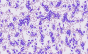
  • Blood samples were received from a nine-year-old male sheep that developed a transient injection site swelling after a second BTV vaccine that resolved in 48hours and then it became anorexic and lethargic for a week. On examination, the sheep had a mildly swollen face, bilateral clear nasal discharge, increased respiratory noise, poor rumen fill and a rectal temperature of 39.6o Haematology initially detected a lymphocyte count of 328.5 x 10⁹/l (RI: 1.6 – 7.5) and follow up examination of the blood smear by a clinical pathologist found increased numbers of atypical round cells, accounting for over 90% of nucleated cells present. These had a moderate amount of pale basophilic cytoplasm and a variably placed irregularly round or occasionally indented or angular nucleus 4-5 times the diameter of an erythrocyte. Occasional examples were bilobed, complex or floriform, chromatin was coarse and cells had one to two small, indistinct nucleoli. Occasional mitotic figures were also seen. The findings were consistent with severe acute leukaemia (Figure 2). Most likely, this was of lymphoid lineage, but myelomonocytic lineage was also possible. The animal was also anaemic and this was probably secondary to bone marrow myelophthisis with neoplastic cells. Lymphoproliferative disease is not uncommon in sheep and is typically of the spontaneous form (as opposed to hereditary or associated with bovine leukaemia virus).

Figure 2: Large numbers of atypical, nucleated round cells in a blood smear from a sheep with leukaemia.
Camelids

  • High strongyle egg counts consistent with parasitic gastroenteritis (1250, 2000 and 2500 epg) were detected in three alpacas from different
  • Patent fasciolosis was diagnosed on routine faecal monitoring in an Rumen fluke eggs were also seen.

Other species

  • Faeces samples were received from an 80-sow farrow-finish indoor pig farm which was experiencing intermittent postweaning scour. Lawsonia intracellularis, the cause of proliferative enteropathy, was detected by PCR in six samples and Brachsypira pilosicoli, the cause of intestinal spirochaetosis, was detected by PCR in six samples.
  • Blood samples were received from a two-year-old pig with a six-day history of red mucous membranes, inappetence, tachycardia and reduced faecal output. Haematology detected a lymphocyte count of 263.1 x 10⁹/l, with the majority of lymphocytes being medium to large in size with small to moderate amounts of basophilic cytoplasm. There were also 3 x 10⁹/l of unclassified large mononuclear nucleated cells with small to moderate amounts of basophilic cytoplasm and nucleoli. These findings were very suspicious of leukaemia.
  • Parasitic gastroenteritis was diagnosed in a reindeer with a strongyle egg count of 1400 epg.
  • A Lohmann Brown hen was euthanised due to coelomic cavity distension and lethargy. On PME, multifocal yellow circular lesions were present throughout the liver and there was an abnormal pedunculated liver lobe mass. There was also a markedly thickened oviduct (salpingitis) and signs of early egg peritonitis in the coelomic cavity. Samples of liver were submitted for histopathology and there was evidence of multifocal hepatic lipidosis. There were also occasional accumulations of fibrin-like material expanding sinusoids multifocally. Hepatic lipidosis can cause haemorrhagic diatheses in affected birds which are often female and in fat body condition. There is evidence to suggest that repeated haemorrhages are a feature of this condition, ultimately resulting in fatal haemorrhage and hepatic rupture. Haemorrhages may have led to the lethargy reported in this case.
  • Mycoplasma gallisepticum infection was confirmed on PCR testing of a swab taken from the eye and nostril of a chicken.
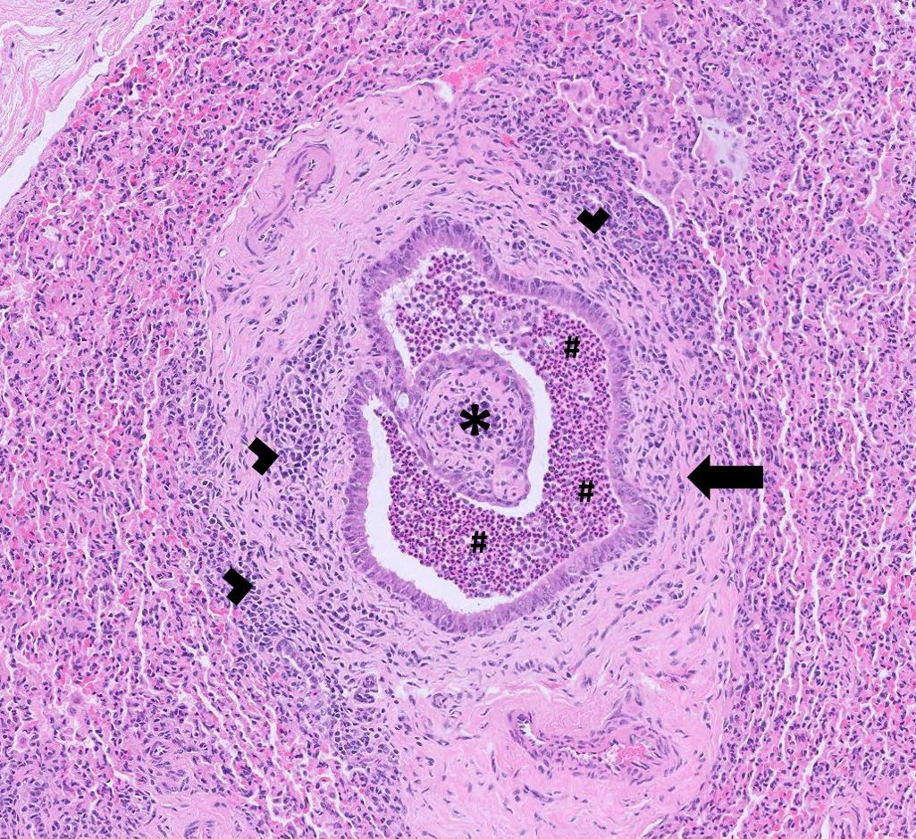
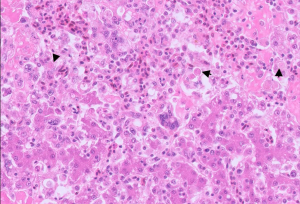
  • Post-mortem tissues wee received from two chickens after four out of a total of eight birds had died suddenly, with no clinical signs At PME, lesions were seen on the liver that were suspicious of histomoniasis and this was confirmed on histopathology, with the presence of multifocal to coalescing, necrotising and granulomatous hepatitis with intralesional protozoal trophozoites (Figure 3)

Figure 3: Trophozoites (arrows) of Histomonas meleagridis in the liver of a chicken with histomoniasis.

Deficiencies and toxicities

  • Tissues were received from a ewe which had been euthanised on welfare ground after showing acute onset signs of jaundice, recumbency, harsh lung sounds and shivering. Histological examination of spleen was unremarkable and in the lung tissue there was oedema and congestion with small multifocal The most marked changes were in the liver tissue which revealed severe chronic hepatopathy characterised by marked bridging fibrosis, nodular regeneration, ductular reaction, ongoing hepatocellular degeneration, and multifocal hepatocellular necrosis. The presence of abundant intracytoplasmic orange granular pigment, presumed to represent copper, together with the overall pattern of hepatic injury, was considered highly suggestive of chronic copper toxicosis with subsequent acute hepatocellular injury and copper release, resulting in a haemolytic crisis. Chronic copper poisoning is a well-recognised condition in sheep and reflects their ability to accumulate substantial quantities of copper within hepatocytes over prolonged periods without overt clinical signs. Clinical disease typically develops only once hepatic storage capacity is exceeded or following a triggering event that results in sudden hepatocellular injury and release of large amounts of copper into circulation. Excessive dietary copper intake is the most common underlying cause and may result from contaminated feed, pasture, water sources, or inappropriate mineral supplementation. In addition, deficiencies of molybdenum, sulphur, or iron may enhance intestinal copper absorption and predispose animals to excessive hepatic copper accumulation.
  • Pregnancy toxaemia was suspected to have resulted in elevated NEFA levels of 863 and 1783µmol/l (RI = 0-600µmol/l) in two cows which had been diagnosed as having ketosis on an in-house test. They were close to calving, and one animal was recumbent and the other still standing but weak. Recent drought conditions resulting in a diet with insufficient supply of energy and nutrients was suspected as the underlying cause.
  • Fatty liver disease was suspected in a cow which was ten days into lactation and had undergone surgery for left displaced abomasum three days The cow had been eating and poorly post operatively and had reduced rumination. In house testing had revealed some evidence of ketosis. Liver enzymes GLDH and GGT were raised at 152U/L (RI= 0-25U/L and 62U/L (RI= 0-50U/L) respectively. There are numerous possible causes of raised liver enzymes (e.g hepatic abscesses, endotoxaemia, liver flukes, plant toxicosis, copper toxicosis and mycotoxicosis but in this case hepatic lipidosis was thought to be a likely explanation, though not all cows with fatty liver disease will have significantly raised GLDH and GGT levels.
  • Two of a group of six cattle were found to have elevated blood lead The screening tests were carried out after high lead levels were found in tissues from an animal in the same group, following testing at a postmortem facility. In the screening submission both animals had blood lead levels between 0.15 and 0.48 umol/l. The Food Standards Agency requires that animals with lead levels within this range may only enter the food chain if presented for sale or to the food chain with food chain information (FCI) which states that offal should be discarded.
  • Evidence of hypocalcaemia (<1mmol/l) and marginal hypomagnesaemia (0.63mmol/l) were measured in ocular fluid from a cow which had died suddenly and from which the sample was taken within one hour of death. Aqueous humour and vitreous humour calcium levels of < 1 mmol/l are suggestive of terminal hypocalcaemia. Vitreous humour magnesium levels of < 0.55 mmol/l are considered useful as diagnostic indicators of hypomagnesaemia for up to 48 hours after death in cattle.
  • Hypocalcaemia was diagnosed in a cow which was down and bloated at around 50 DIM. There was a calcium level of 1.51mmol/l (RI= 2.00.3.00mmol/l) and mildly raised CK and AST levels suggested that a degree of muscle tissue injury had also occurred.
  • There was evidence of hypomagnesaemia in a seven-year-old South Devon cow, which was reported to have ‘done the splits’ raising fears of ligament and nerve damage. The magnesium level on a blood sample was below the reference interval at 58mmol/l (RI 0.7-1.3mmol/l). CK and AST were raised at 775U/l (RI = 0-250U/L) and 154U/L (RI=0-100 U/L) indicating that some very minor muscle damage was present.
  • Iodine and copper deficiency were suspected in a group of five barren Lincoln Red cows which were in poorer condition than the rest of their The group had a low result on the pooled inorganic iodine test of 37ug/l (levels >150ug/l indicate adequate current iodine intake) consistent with low recent iodine intake in the majority of those contributing to the pool. All five animals had evidence of hypocupraemia. As plasma copper concentrations decline, the probability of a copper responsive disease being present increases but hypocupraemia alone is not sufficient evidence to confirm copper deficiency disease. Plasma copper concentrations are usually well below the reference interval (9.0-19.0µmol/l when clinical copper responsive disease occurs, typically less than 6.0 µmol/l.
  • There was evidence of selenium deficiency on analysis of liver tissue from a six-year-old Jersey cow. Postmortem examination had revealed the abomasum was full of sand and an omasum distended with rumen contents, suggestive of an impaction following suspected pica behaviour. The selenium level in liver was 8µmol/kg DM (RI 11.0-22.6µmol/kg DM), confirming likely selenium deficiency and although it could not be certain how exactly this related to the postmortem findings, it is an unusual finding in an adult dairy cow.
  • There was evidence of Vitamin A deficiency in two calves which had serum vitamin A levels of 39µg/l and 79µg/l (RI = 249-500). Clinical history indicated that the calves were not doing well in general, not suckling well at birth and then succumbing to scour and Low vitamin A was observed on several other submissions this month, with a similar history of calves not doing well, having scours or pneumonia with no infectious causes found and increased mortality. Care must be taken in interpretation of Vitamin A results from neonatal calves. Colostrum is the main source of vitamin A in the newborn calf, so if there has been insufficient intake of good quality colostrum, vitamin A levels are also likely to be low. There could be a higher risk of vitamin A deficiency occurring this autumn through to next spring following drought conditions in many areas this summer.

Follow us on Facebook

https://www.facebook.com/Axiomfarmvetlab/

This newsletter is sent by e-mail to each vet practice but if you would like a copy sent to your individual e-mail account please contact us at dsfarm@axiomvetlab.co.uk and we can add you to our circulation list.

Related Posts