Farm September Newsletter 2026

Dedicated farm line – 01626 357776

Follow us on Facebook

https://www.facebook.com/Axiomfarmvetlab/

This newsletter is sent by e-mail to each vet practice but if you would like a copy sent to your individual e-mail account please contact us at dsfarm@axiomvetlab.co.uk and we can add you to our circulation list.

Would you like to send the Axiom farm vets photos of PMEs to allow us to assist with sample selection and diagnosis? Please send photos by WhatsApp to 07944 649 222.

Note that we may wish to include photos sent to this number for the interesting cases section of this newsletter. If you do not wish your photo to be used, please let us know at the time of sending.

Email us with your additional test requests

You may find this is a more efficient way of making requests than phoning the farm team, saving you time in your busy day. Our farm team also find it a more efficient way of dealing with your requests.
The email address for test requests is: dsfarm@axiomvetlab.co.uk 

Bluetongue testing guidance

Please note that, due to SAPO regulations, we cannot accept samples from animals showing signs suspicious of Bluetongue infection, being tested for Bluetongue or that have recently tested positive for Bluetongue. If such samples are received, they will have to be destroyed. We can accept samples from herds or flocks which are currently undergoing testing for BTV or are showing signs consistent with BTV infection provided there is no suspicion of bluetongue virus infection in the individual being tested. We are happy to discuss cases over the phone before submission of samples. Clinical suspicion of BTV infection must be reported to APHA.  For the latest information and updates on BTV in the UK please visit the following link:

Bluetongue: latest situation – GOV.UK

Sheep scab serology

We wanted to make clients aware of a temporary change to sheep scab serology testing. Following recent submissions, we have seen a small number of results that did not seem to fit with the clinical history, including positive results from flocks with no clinical signs of sheep scab and/or where animals were being tested for screening purposes. We are working closely and constructively with the referral laboratory to review these findings, consider possible explanations and agree the safest and most reliable way forward. While this work is ongoing, and as this is currently the only laboratory offering this testing, we have made the precautionary decision to pause referrals for sheep scab serology through Axiom.

We appreciate that this may be inconvenient and are sorry for any disruption it causes. Because a positive sheep scab result can have significant implications for clients, we feel it is important to take a careful approach while we continue to work with the referral laboratory to understand the issue fully and identify a solution. We hope to be able to resume this service as soon as possible and will keep clients updated as soon as further information is available.

Increase in Haematological abnormalities

We appear to be seeing an increase in the number of ruminants with abnormal haematology findings in recent months. Thrombocytopaenias, lymphopaenias and/or lymphocytosis have been more commonly seen than usual. It is possible that these changes are due to BTV infection. One study where sheep were experimentally infected with BTV2 found that leukopaenia occurred from two to seven days post infection (dpi), lymphopaenia was common at 3 dpi, marked thrombocytopaenia occurred from 2-14 dpi, PCV and haemoglobin decreased 3-21 dpi and at days 11-14 post infection leukocytosis due to lymphocytosis was seen. Haematological and biochemical alterations in native sheep experimentally infected with bluetongue virus serotype-2. Khorajiya JH et al. Indian Journal of Animal Sciences 89 (1): 3–8, January 2019. Another study involving infection with BTV 1,

9 or 23 also found evidence of thrombocytopaenia and lymphopaenia with a nadir at 8-11 dpi. Bluetongue virus infection in sheep: haematological changes and detection by polymerase chain reaction. K A McColl , A R Gould. Aust Vet J. 1994 Apr;71(4):97-101. Unfortunately, if animals are suspected of having BTV we are unable to receive samples from them. See later in the newsletter for some interesting “bleeding calf cases”.

Interesting cases for July and August

Cattle
Abortions/mastitis

  • Fixed tissues were received from a six-month gestation aborted male shorthorn foetus. Autolysis hampered much of the histopathological examination but within the lung and liver small numbers of neutrophils were observed which could be suggestive of bacterial infection. Within the foetal liver there was evidence of biliary stasis. In the foetus the placenta assists with bile excretion and so biliary stasis can be seen with placental insufficiency. Possible causes of placental insufficiency can include placentitis (e.g. due to an infectious cause), poor uterine perfusion or systemic illness in the dam, multiple foetuses, poor maternal nutrition, torsion or compression of the umbilical cord, toxins such as mycotoxins, and heat stress. Light, pure growths of Aeromonas spp. were isolated in culture of the foetal stomach contents and this finding along with the presence of inflammatory cells and biliary stasis on histopathology was considered of potential significance as a cause of this sporadic abortion.
  • Profuse growths of Lelliottia spp. were cultured from the stomach contents of a seventh month gestation dairy calf foetus. Lelliottia spp. is a member of the Enterobacteriacae and could be a potential cause of abortion due to opportunistic invasion of the gravid uterus.
  • Pasteurella multocida was isolated in cultures of foetal stomach content in one case of abortion in a beef cow. multocida is a cause of sporadic abortions in cattle typically occurring by haematogenous spread e.g from the respiratory tract or septicaemia.
  • There were at least eight cases over July and August in which Salmonella was isolated from foetal stomach contents, in which the isolates were identified as Salmonella Dublin.
  • A profuse and pure growth of Helcococcus ovis was isolated from a mastitic milk sample from a dairy cow. This isolate is rarely reported as a cause of bovine mastitis but has also been found in other sites in farm animals e.g abscesses, metritis, endometritis and bronchopneumonia.
  • Klebsiella pneumoniae was isolated from milk samples from two cows on a dairy unit which was experiencing an increase in mastitis cases after drying-off. Outbreaks of Klebsiella mastitis have been associated with sawdust bedding. Klebsiella can produce a severe toxic mastitis.
  • Cytology of material expressed from the teat of a two-month-old male calf identified marked neutrophilic inflammation, consistent with bacterial mastitis. No bacterial culture of the material was carried out. ‘Summer mastitis’ typically involving pyogenes is one of the most common forms of mastitis in male animals outdoors. Cross-suckling can also occur in male calves housed in groups and this can predispose them to mastitis by introducing bacteria directly into the teat canal.
  •  

Respiratory disease

  • IBR virus was detected by PCR in a pool of three ocular swabs from seven-month-old mixed breed calves which were showing signs of pyrexia, coughing oculonasal discharge, chemosis and On another farm IBR was also diagnosed by PCR on a nasopharyngeal swab in an adult cow with respiratory signs and pyrexia. IBR was detected by PCR in at least eleven other cases in July and August.
  • Fixed samples of lung tissue from a five-month-old calf were submitted to investigate the cause of respiratory signs and increased mortality. Unfortunately, marked to extreme autolysis and postmortem bacterial contamination hampered histopathological examination, though in some better-preserved areas there was evidence of moderate fibrinosuppurative bronchopneumonia and nematode larvae were Parasitic pneumonia due to Dictyocaulus viviparus with secondary bacterial infection was considered the most likely cause of the lung lesions in this case. There were multiple other cases of lungworm diagnosed through July and August. In one case half of the cows in a small dairy herd of twenty animals were affected with coughing and showed some degree of milk drop with two animals appearing clinically sick.
  • Mannheimia haemolytica was detected by PCR of a lung swab from an Aberdeen Angus cross calf with evidence of severe pleuropneumonia on postmortem examination.
  • A postmortem was carried out on farm of a ten-week-old calf which had appeared to have died suddenly. The animal had been out at pasture with its dam since the age of two Extensive consolidation of lung tissue with abscesses throughout was observed. Trueperella pyogenes, a common secondary pathogen found in abscessated lungs was cultured from a lung swab and PCR testing detected Mycoplasma bovis, Histophilus somni and Pasteurella multocida, all of which could be considered of significance, usually acting as secondary pathogens.
  • A five-month-old calf presented with signs of coughing and respiratory distress. There was little response to treatment with long acting oxytetracycline and flunixin and the animal died within three days of the onset of clinical signs. Samples of fresh and fixed lung tissue were received. Pasteurella multocida was detected by PCR testing of the fresh tissue. Histopathology revealed that there were severe, chronic and ongoing changes in the lung consistent with a case of enzootic calf pneumonia/ bovine respiratory disease. Bronchiolitis fibrosa obliterans pointed to a severe necrotising bronchiolitis suggestive of a previous pneumotropic viral infection such as RSV, PI or IBR. However, PCR testing was unrewarding which may have been due to the chronic nature of the lesions. Evidence of alveolar septal necrosis with hyaline membrane formation raised the possibility of this being a case of BIP (bronchopneumonia with interstitial pneumonia). BIP is considered a distinct condition where chronic cranioventral bronchopneumonia and inflammation increases sensitivity to 3-methylindole (or another pneumotoxin), leading to acute caudal interstitial lung disease. However, to make this diagnosis correlation with gross pathology would be required, which was not possible in this case.
  • Diffuse Alveolar Damage (DAD), also known as interstitial pneumonia was the likely cause of peracute illness and death in a pre-weaned Limousin cross DAD represents a non-specific histopathological pattern of acute lung injury resulting from primary insult to the alveolar epithelium and/or vascular endothelium. As such, it is associated with a wide range of aetiologies in cattle, which can broadly be divided into toxic, infectious, and systemic causes. In cattle, one of the most recognised causes is exposure to pneumotoxic compounds, particularly 3 methylindole (fog fever) however, this is less common in pre-weaned animals. Other toxic insults include plant-associated pneumotoxins (e.g. perilla mint) and dietary or environmental toxins. Infectious agents may also result in DAD, either through direct injury to alveolar structures or indirectly via inflammation (cytokine storms); these include respiratory viruses (notably bovine respiratory syncytial virus), parasitic infections such as lungworm, and septicaemic processes. Given this broad range of potential causes, interpretation of DAD requires integration with clinical history, management factors, and ancillary testing, as the lesion itself is not pathognomonic for a specific aetiology. There was a similar case on another farm where a heifer presented with pyrexia, cyanosis and acute respiratory distress. Six other animals in the group were also affected. PCR testing of lung tissue did not detect any infectious agent but there was evidence of DAD/ interstitial pneumonia on histopathology. In this case the animals had been recently moved during hot weather, and it was considered that heat stress may have predisposed the animals to respiratory disease.

Gastrointestinal disease

  • There were numerous cases of coccidiosis and suspected coccidiosis in calves in July and August. Eimeria zuernii, one of the two main pathogenic species of coccidia in cattle, was found to make up 88% of the oocysts present in a pooled faecal sample with a coccidial oocyst count of 20,500opg from a group of three to four-month-old scouring calves, confirming a diagnosis of coccidiosis in that case. Coccidiosis was suspected to be the cause of severe haemorrhagic scour in a group of Aberdeen Angus calves. The coccidial oocyst count was >50,000opg. Eimeria species identification was not carried out, though very high counts such as this are usually significant in cattle. Salmonella cultures were negative. In another case fixed tissues including sections of small intestine were received from a post weaned Holstein-Friesian dairy calf. Examination of the gut tissue revealed a chronic eosinophilic and granulomatous enteritis with intralesional coccidial structures suggestive of mucosal damage due to coccidiosis, though no further details regarding the clinical signs were given.
  • Coronavirus was detected in at least four cases of neonatal calf diarrhoea and in two of these Cryptosporidium antigen was also detected.
  • E.coli F5 (K99) antigen was detected in the intestinal contents of a calf which had appeared healthy at birth but became ill and died at less than twenty-four hours of age. Yellow fibrin strands were observed in the peritoneal cavity at postmortem examination. Antigen testing for rotavirus was also positive in this case, though clinical signs attributed to rotavirus do not usually occur in calves before around five days of age. Therefore, the enterotoxigenic E.coli was the most significant finding in this case. There were at least five other cases of E.coli F5 (K99) associated enteritis in neonatal calves in July and August.
  • There were multiple cases of gastrointestinal disease in adults and youngstock related to Salmonella In one submission Salmonella Typhimurium was isolated in a sample from one of two pyrexic dairy cows. There was a history of sick calves and recent abortions on farm.
  • A high strongyle egg count of 700epg was recorded in a faecal sample taken from one of three collapsed calves in a group of 75 three to four-month-old Holstein-Friesian heifers on Watery, dark scour was observed in the affected animals and this along with the high faecal worm egg count was indicative of parasitic gastroenteritis. No coccidial oocysts were observed in the sample and Salmonella cultures were negative making these differential diagnoses less likely, though it should be considered that disease due to coccidiosis can occur in the prepatent stages and that shedding of Salmonella can be intermittent.
  • Pepsinogen levels were significantly raised in two Wagyu fattening animals which were not thriving and were found to have each lost 30kg at weighing. One of the two was also scouring. The anthelmintic treatment history was not given but no eggs were observed on individual faecal egg Pepsinogen levels were raised at 4.83iu/l and 3.97iu/l (RI = 0.00-1.50). These levels are considered indicative of significant abomasal damage due to parasitism which was likely to be associated with the reported clinical signs.
  • Mycobacterium avium paratuberculosis (MAP) was detected in the faeces of an adult cow at a low Ct value of 24, indicative of heavy shedding of the bacterium and consistent with a diagnosis of Johne’s disease. In addition to the positive Johnes’ PCR result, Bovine coronavirus (BCov) was detected using the bovine coronavirus antigen test on faeces and this result would be considered diagnostic of winter dysentery. The coronavirus lateral immunochromatography antigen test is not usually recommended for detection of winter dysentery as it is thought to be of insufficient sensitivity to detect the disease in adult cattle. PCR testing or paired serology are the preferred diagnostic tests for this condition. There were numerous other cases of Johne’s disease diagnosed including a twenty-month -old British Blue cross animal which had been losing condition and scouring over the previous two months and an Aberdeen Angus bull which had signs of severe wasting and scour for one month.

Skin and eye disease

  • Ringworm (Dermatophytosis) was diagnosed by PCR on a hair pluck from one of five dairy heifers with skin The infection had spread through the herd and most had recovered but these animals remained affected.
  • Corynebacterium pseudotuberculosis, the causative agent of caseous lymphadenitis was isolated from a swab from an unknown site on an adult No other history was available but CLA is occasionally known to occur in cattle.
  • There were at least nine submissions in which Moraxella bovoculi was isolated in cultures from cattle showing signs of infectious bovine keratoconjunctivitis (IBK), also known as New Forest eye or pinkeye. In several of these cases the bacterium was isolated from more than one animal in the same Staphylococcus aureus was isolated in addition to M.bovoculi on one sample and this was a possible significant pathogen acting as an opportunist and contributing to the eye lesions in that animal. M. bovoculi is commonly isolated in cases of IBK in cattle but can also be found in healthy animals. In another case of suspected IBK three eye swabs from affected animals were cultured and Pantoea spp. were isolated from all three. Pantoea spp. are commonly found in the environment but could be acting as opportunists in this case e.g following irritation from dust or flies. In a third case of IBK, Rothia nasimurium was isolated from an eye swab. It was noted that in one study by Gafen et al (2023) Rothia nasimurium was significantly more abundant in IBK eyes than in normal eyes so it could be an opportunistic pathogen in this case.
  • Penile fibropapilloma was diagnosed by histopathology of a tissue biopsy from a Beef Shorthorn bull. Fibropapillomas are hyperplastic lesions caused by papillomavirus infection. Penile fibropapillomas in bulls are most often diagnosed at one to two years of age. They can spontaneously resolve, but this occurs after a variable time period, and recurrence can often be a problem.

Systemic and miscellaneous disease

  • Notable cases of BVD viraemia included a two-day-old bull calf which was dull, had reduced mentation and failed to rise after The calf tested positive for BVD by PCR on a blood sample. In another case a poorly thriven twenty-month-old Aberdeen Angus cross bullock tested positive in the BVD antigen ELISA raising a strong suspicion that it was a BVD persistently infected animal. In a third submission a bought-in yearling heifer showed signs of poor appetite and was failing to thrive. It had a high positive result for BVD antigen in the ELISA and it was suspected that this animal was also likely to be a BVD PI.
  • Blood parasites associated with erythrocytes and with an appearance consistent with Babesia sp. were seen in one out of three blood smears submitted from adult dairy cattle in south west Scotland with signs of milk drop, haematuria and anaemia. Babesiosis was also diagnosed on examination of blood smears from two dairy heifers in North Somerset showing signs of red urine, but which were otherwise well in themselves. In another submission from a farm in Cornwall an adult dairy cow with milk drop, blood in the urine and pyrexia tested positive for Babesia sp. by PCR testing of an EDTA blood sample. Babesia sp. were detected by PCR testing of a blood sample from a six-year-old Dexter cow in Gloucestershire which became jaundiced, anaemic and pyrexic around one month after calving. In a fifth case in mid-Wales a yearling heifer with respiratory signs and jaundice had both evidence of Anaplasma phagocytophilum inclusions in the neutrophils on the blood smear, confirming Tick borne fever and parasites consistent with Babesia sp. within the erythrocytes indicating concurrent
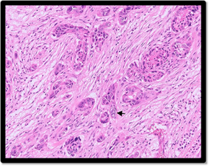
  • Renal carcinoma was diagnosed in a 16-year-old, male, castrated miniature Zebu which was found dead At postmortem examination the right kidney appeared misshapen and around 75% of the tissue was very firm and partially adhered to the peritoneum. Purulent material was observed on incision into the renal pelvis. Histopathology of a section of the kidney revealed that around 60% of the section consisted of an unencapsulated, poorly differentiated collection of neoplastic cells. Similar cells were observed in a section of nearby lymph node. Taking into account the gross description of the kidney and the histopathological appearance, it was considered that this tumour was most likely a malignant renal carcinoma with spread into the local lymph node.

Figure 1: Clusters of neoplastic epithelial cells in the kidney of a zebu with suspected renal carcinoma. Arrow indicates an atypical cell with karyomegaly (large nucleus).
  • Fixed tissue samples consisting of sections of oesophagus and lung were submitted from one animal from a group of Jersey cross calves with signs of malaise and poor performance associated with some mortality. Histopathological examination revealed marked lesions with evidence of severe ulcerative oesophagitis and bacterial bronchopneumonia. There was evidence of bacterial colonisation of the lesions in the Such lesions are often associated with mucosal trauma, including injury secondary to coarse or abrasive forage, foreign material, or iatrogenic damage (e.g. administration of oral medications or dosing equipment), although other causes of mucosal injury could also be considered (e.g uraemic syndrome). It was commented that summer scour syndrome (SSS) of dairy calves cases can sometimes have oesophagitis. SSS usually occurs within a few weeks of turnout and is believed to be due to rumen dysbiosis due to too rapid a change in ration. It can be more common on lush grass and it is also possible that the high oil levels in the grass contribute.
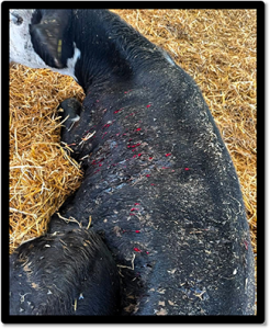
  • Pre and postmortem samples were received from a four-month-old Holstein dairy heifer calf that showed signs of acute haemorrhage with multiple small wounds on the skin and torso which failed to clot (Figure 2). A prolonged clotting time was recorded during the clinical Petechial haemorrhages were visible on the hairless skin, vulval membranes, in the conjunctiva/sclera and on the internal organs at postmortem. Unclotted blood was present in the abdominal cavity. Haematology revealed severe anaemia with a PCV of 9% (RI=24-40%) and the platelet count was 16 x 10^9/L (RI= 200-800 x10^9) indicating marked thrombocytopaenia, which could explain the spontaneous haemorrhages and petechiae of mucous membranes. On histopathology there was evidence of bacterial bronchopneumonia and interstitial nephritis, most likely indicative of a past bacteraemia (e.g colisepticaemia). The findings considered most significant were seen in bone marrow tissue. There was a marked hyperplasia in the bone marrow suggesting the haemorrhage had been occurring for longer than previously assumed (at least 24 hours) (Figure 3). The cause of the bleeding / lack of clotting was not possible to determine from histopathology. Initial differentials which were considered included DIC, anticoagulant rodenticide toxicity, inherited deficiency of coagulation factors and ingestion of mouldy sweet clover. A similar case of anaemia (PCV 15%) with thrombocytopaenia (Platelets 10×10^9/l ) with haemorrhage from multiple sites was seen later in August and in that case, the heifer, which was the only animal affected died soon a short time after examination (Figure 1). The unusual nature of these cases triggered further discussion and a possible increase in submissions involving thrombocytopaenia in farm animalsover previous weeks had been observed. There is a possible link of thrombocytopaenia with BTV infection, and this is being further investigated in collaboration with APHA. Thrombocytopaenia, along with other haematological abnormalities, has been documented in previous Bluetongue cases. Van den Brink KMJA, Santman-Berends IMGA, Harkema L, Scherpenzeel CGM, Dijkstra E, Bisschop PIH et al. Bluetongue virus serotype 3 in ruminants in the Netherlands: Clinical signs,seroprevalence and pathological findings. Vet Rec. 2024;e4533. Three cases of bleeding calves with haemorrhaging from the skin linked to BTV8 and a single case with BTV3 in calves aged 1-2 weeks old have been reported in France.

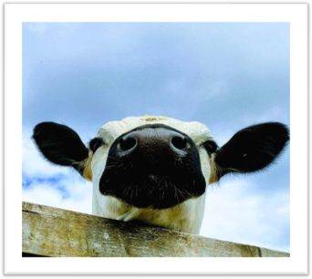
Figure 2 : Down heifer with signs of haemorrhage from multiple sites with failure to clot. (Photo Sophie Goodings, Axe Valley Large Animal Vets)
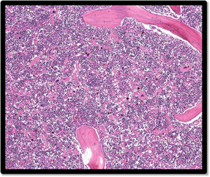
Figure 3: Bone marrow from a bleeding calf showing near 100% cellularity, with hyperplasia of the erythroid cell line.

  • Clostridium botulinum C/D toxin was detected by ELISA testing of small intestinal contents taken at postmortem examination of a yearling heifer which died after a short duration of The finding was suggestive of a diagnosis of death due to botulism, though in autolytic carcases there can be a risk of postmortem formation of botulinum toxin. It was advised to consider any known risk factors and clinical signs and to contact APHA for advice relating to potential food safety concerns.
  • A two-day-old heifer calf which was a twin to a bull calf was tested in the freemartin genetic test on an EDTA blood sample and male DNA was detected, confirming the animal was a freemartin. This condition affects about 90% of female calves born with a male twin, due to the fusing of the two placentas and sharing of blood supply and hormones in utero.
  •  

Sheep and goats

  • In a hefted, closed hill flock of around 700 Welsh ewes, 80 to100 ewes, scanned in lamb at the end of January, had not Ticks were prevalent on the open hill shared with other grazers and the ewes were poorer than normal. but were treated for ticks regularly. All six sampled sheep had strongly seroconverted on Tick Borne Fever serology, suggesting recent exposure to Anaplasma phagocytophilum, and also on Toxoplasma gondii serology. Two were also seropositive on Border disease serology.
  • Further cases of seroconversion to Toxoplasma gondii included a 190 ewe self-replacing flock that started aborting at lambing time, with aborted lambs either fresh dead or dead for several Many living lambs were weak and ewes were also dying and many were barren at lambing. Eight of the ten ewes sampled had seroconverted strongly to T. gondii.
  • Further cases of Mannheimia haemolytica and Staphylococcus aureus mastitis were reported in These are the two most common causes of severe mastitis in sheep. A profuse growth of a Trueperella sp. was isolated from another ewe with mastitis and this is also a known cause of mastitis in sheep as well as in cattle.

Respiratory disease

  • Acute fibrinous suppurative bacterial bronchopneumonia was diagnosed on histopathology of lung samples from a thin adult sheep, most likely due to descending bacterial infection. Given the apparently acute inflammatory process without convincing evidence of chronicity, it was uncertain how these changes related to the animal’s poor body condition. Culture would have been required for further bacterial identification, but the appearance was suggestive of involvement of the Pasteurellaceae.
  • Sudden death with no premonitory clinical signs was reported in three lambs in one flock and postmortem samples were received, with lung changes on PME reported to be characteristic of Pasteurella Consistent with this, the main histological findings were a marked chronic suppurative bronchopneumonia including intralesional bacteria. In addition, some of the changes were suggestive of ‘atypical pneumonia’ and the underlying cause may have been multifactorial, possibly involving viral and mycoplasma agents as well as bacteria such as the Pasteurellaceae (which would fit with the gram-negative bacteria seen in lesions). However, other features of ovine enzootic pneumonia/atypical pneumonia were absent, such as peribronchiolar lymphoid cuffing and airway epithelial hyperplasia.
  • Post-mortem tissues were received from a lamb, one of 5% of lambs lost in a group that had received Heptavac at three weeks Serous nasal discharge was present in approximately 50% of the group and some were also coughing. Consistent with the reported gross findings on PME there were extensive areas of necrosis and abscessation. These could have been the result of chronic pneumonia (if the reported lesions were in the cranioventral lung) or embolic spread from a focus of inflammation elsewhere (which was potentially more likely if abscesses were throughout both lungs). There was also evidence of a more acute terminal bacterial pneumonia most consistent with Mannheimia haemolytica, although this could also have been caused by Pasteurella multocida or Histophilus somni which were detected on PCR of lung. PI3 virus was also detected by PCR and this may have been the cause of observed airway damage. There was also a minimal lymphoplasmacytic infiltrate which may have been due to ongoing antigenic stimulation or Mycoplasma ovipneumoniae involvement.
  • Post-mortem tissues were received from a ewe with an ongoing history of weight loss and bilateral nasal discharge. Consolidation had been seen on lung scanning and there was no improvement on worming, the ewe subsequently dying. Sheep had been bought in from a farm that had since been diagnosed with an OPA outbreak and lung histopathology confirmed that this was also a case of OPA. A second case was diagnosed in a shearling Texel ewe with a one-week history of lethargy and pyrexia (temp 3oC). In this case there was also evidence of a mild chronic bacterial infection which may have explained the reported pyrexia. Perhaps the most notable case was in a reportedly five-month-old lamb which had died suddenly after a bout of mild coughing and sneezing. The lungs appeared consolidated and hyperinflated on PME. There was evidence of OPA with secondary necrosuppurative pneumonia which would have accounted for the sudden death. Likely pathogens to have been involved would have included Mannheimia haemolytica, Bibersteinia trehalosi and Pasteurella multocida. Infection normally occurs at a young age with progressive neoplastic transformation of pulmonary tissue and onset of clinical signs normally from two to three years. However, we do see cases in much younger sheep.

Gastrointestinal disease

  • A coccidial oocyst count of greater than 50,000 opg was detected in four-month-old scouring lambs, of which 76% were of the pathogenic species, Eimeria ovinoidalis. Although coccidiosis is typically seen in lambs under two months of age, it was suspected that the count was significant in this case. There may have been a lack of challenge at a younger age. No strongyle eggs were seen and Salmonella was not
  • A male goat developed sudden onset watery scour with mucus and red The goat deteriorated rapidly and bloated before dying. PME detected extensive pulmonary oedema, ulceration and inflammation of the large intestine. Clostridium perfringens epsilon toxin was detected by ELISA of small intestinal contents, supportive of a diagnosis of Type D clostridial enterotoxaemia. C. perfringens epsilon toxin was also detected in faeces from a male Golden Guernsey goat that got in with some sheep and was suspected to have gorged on sheep food. The animal deteriorated rapidly over the course of a day and was in a lot of pain.
  • Multiple cases of parasitic gastroenteritis were diagnosed in sheep and This included several cases of suspected haemonchosis in sheep flocks and some goat herds with strongyle egg counts in excess of 1000 epg, some of which were confirmed on Ovacyte microscopy, with pale mucous membranes/ anaemia and bottle jaw reported in some animals.
  • Raised pepsinogen levels consistent with significant abomasal parasitism were reported in sheep and goats, including a 16-month-old Suffolk ram with ill thrift and poor muscling despite concentrate and fodder
  • Nematodirosis and coccidiosis appeared to have been contributory factors in the death of twin six-week-old lambs that died after developing scour. A faecal sample from one lamb had a Nematodirus battus egg count of 200 epg and a coccidial oocyst count of 7550 opg, of which 44% were identified as the pathogenic Eimeria species, ovinoidalis.
  • Cases of fasciolosis were diagnosed in sheep on coproantigen ELISA and
  • Cases of Johne’s disease diagnosed on serology and faecal PCR included a four-year-old, ill thrifty male goat with hind limb ataxia and a ewe with a waxing and waning history of weight loss and recent scour. In one flock, three thin two-year-old ewes had single lambs this year and were BCS 1.5 in a flock where first time lambers were average BCS 2.5 and older ewes 3 – 3.5. Two ewes had been diagnosed with Johnes 18 months ago and, of the three, one was seropositive and two were faecal PCR positive.
  • Salmonella 61:k:1,5,7 was isolated from one of three faeces samples from sheep with presumed This is the most common serotype found in sheep and is host adapted.

Skin and eye disease

  • Dermatophilus congolensis was detected on PCR testing of scab material from one of approximately 20% of all yearling sheep exhibiting pruritus in a Blue faced Leicester flock, confirming it as a case of dermatophilosis.
  • Ringworm was confirmed on PCR as the cause of round, hairless skin lesions on a Texel sheep. Unfortunately, as culture was not successful, it was not possible to determine which species was implicated. In sheep it is usually either Trichophyton verrucosum or Microsporum canis.
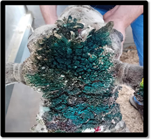
  • Tissues were received from a post-weaned, four to six-month-old ewe lamb that had large growths across the face, back and feet that had appeared rapidly in the last month (Figure 4). Histopathology was consistent with severe, chronic, hyperplastic dermatitis with ballooning degeneration and intralesional eosinophilic intracytoplasmic inclusion bodies typical of parapox (orf) infection. Trauma (e.g. local abrasion from rough grazing) may have predisposed to It was advised that confirmation through orf PCR testing should be considered. Orf was diagnosed in at least one other flock on histopathology and at least three other flocks on PCR testing of scab material. In one of the PCR positive cases, proliferative lesions of readily removable horn-like material were present on the top of head and down the back of the neck of a two-year-old Beltex ram which was the only animal affected.

Figure 4: Orf lesions on the head and neck of a Beltex ram (Photo: Claire Davies, the Fold Farm Vets)
  • Tissue samples were received from a sheep with a lump in the skin on the brisket. Histopathology detected a proliferation of neoplastic polygonal epithelial cells arranged in cords, trabeculae, and islands extending from the surface epidermis, breaching the basement membrane and infiltrating the underlying dermis. This was most consistent with squamous cell carcinoma, although a malignant hair follicle tumour was also a possible differential (e.g. malignant trichoepithelioma). Squamous cell carcinoma in the skin is usually locally invasive, but uncommonly metastatic until late in the course of disease. When spread occurs it is usually to the local lymph nodes in the first instance. This is a relatively common neoplasm of adult to older sheep, most often associated with ultraviolet light damage and are therefore most often seen in pink skin or skin that is lightly wooled. Lesions can be single or multiple, and are most seen on the muzzle, lips, eyelids, pinnae, perineum and vulva. A second case was diagnosed after histopathology of a sample from an area of apparent proud flesh in a crossbred ewe.
  • Staphylococcus aureus was isolated in culture from a swab taken from an area of encrusted skin in a three-year-old male Saanan goat with ocular discharge and nasal S. aureus is a cause of periorbital eczema, which is usually initiated by skin trauma, e.g. banging heads, resulting in skin abrasions and entry of opportunists. S. aureus was also isolated from ulcerated skin lesions on two sheep on a smallholding.
  • Mycoplasma conjunctivae infection was diagnosed on PCR testing of a plain ocular swab in a sheep with an unresponsive eye infection. This is the most common cause of infectious keratoconjunctivitis (‘pinkeye’) in sheep and infections regularly recur due to the difficulty in eradicating the bacterium from deep within the conjunctival sac.

Systemic and miscellaneous disease

  •  Among the cases of Border disease viraemia was a hairy, ill thriven lamb in a flock which had a few cases of BDV viraemia every year.
  • Seroconversion to Maedi visna was reported in several flocks, including 4/6 ewes in poor body condition, 74/114 sheep undergoing screening in a second flock and 7/27 commercial ewes intended as recipients for pedigree embryo transfer in a third flock.
  • Recent cases of CLA confirmed on aerobic culture included a male goat with reoccurring abscess along the There was no evidence of lymph node involvement and it was originally thought to be a tooth root issue. CLA exposure was also confirmed on serology in several flocks and a Golden Guernsey herd.
  • A transport swab was received from a submandibular abscess which had burst in one of several Texel cross sheep with abscesses. An Actinobacillus was isolated. Although this could not be speciated on MALDITOF, it was likely to be A. lignieresii, consistent with this being a case of cutaneous actinobacillosis. The bacterium usually gains entry through abrasions/wounds in the skin. There was no evidence of CLA.
  • Trueperella pyogenes was isolated from an abscess on the thorax of an 18-month-old Suffolk tup and from a suspected CLA abscess in a three-year-old Lleyn T. pyogenes together with Bacteroides pyogenes was isolated from one of multiple cutaneous abscesses on the brisket and prescapular region of a ewe. Both are bacteria capable of causing pyogenic infections.
  • Post-mortem tissues were received from a gimmer, which had been found recumbent, star gazing and seizuring before death. A few other cases had died despite treatment with The affected sheep were at grass with no access to silage or cake. The lesion distribution in the brain was consistent with CCN and this would have accounted for the clinical signs and death of this gimmer. The most likely causes, considering the distribution, were thiamine deficiency (the severity of the lesions could account for the lack of response to thiamine) or sulphate toxicity. The former is commonly associated with recent changes in diet resulting in rumen dysbiosis and the production of thiaminase with subsequent vitamin B1 deficiency. Sulphate toxicity is associated with excess intake either through concentrate food and/or water. Chronic lead toxicity can also cause similar brain changes, and it was advised that consideration should be given as whether there had been any possible access to a source. A few eosinophils were also seen within inflammation in the meninges and, in pigs, these are seen with indirect salt toxicity, and so water deprivation was also a possible cause for the brain changes. A second case was reported in a four-year-old, Ryeland ewe, originally suspected of having meningitis and that had its neck extended and was paddling at the time of euthanasia.
  • MCF was diagnosed in a goat by OvHV-2 PCR testing of a heparin blood No clinical history was provided. Clinical disease due to MCF is rare in goats but cases have been reported in the UK, presenting signs including pyrexia, ataxia, muscle tremors and corneal opacity.
  • Two of this year’s lambs were found dead in a flock and a third lamb was found in the field obtunded, with reduced menace reflex and some nasal discharge but no mouth or foot lesions. Histopathology of brain detected peracute/acute vasculopathy suggestive of epsilon toxicosis associated with Clostridium perfringens enterotoxaemia type D (pulpy kidney). This is normally associated with increased amounts of carbohydrate entering the small intestine resulting in clostridial overgrowth and toxin Epsilon toxin targets blood vessels in several organs including the brain where there is characteristic vasculopathy with perivascular hyaline droplet formation. Vaccination is normally successful at reducing morbidity and mortality and the detection of toxin within the distal small intestine contents is also useful in establishing a diagnosis.
  • Evidence of exposure to Anaplasma phagocytophilum, the cause of Tick-Borne Fever, was seen in flocks in Dorset, Devon and Northumberland. In one case, a sheep found recumbent and covered in ticks had seroconverted strongly to phagocytophilum suggesting recent exposure.
  • Calcium carbonate crystalluria was diagnosed in a 15-year-old female pygmy goat that was straining to urinate and urinating This may be seen with grazing leguminous plants, e.g. clovers, or legume or lucerne hays. Calcium oxalate crystalluria was diagnosed in a second pygmy goat, an adult male in a second herd. There is believed to be a higher risk of this occurring in animals on high calcium or high oxalate diets (latter can be found in some plants e.g. rhubarb leaves, sugar beet leaves, spinach, purslane). Crystals may also be found in healthy animals but when present in high numbers they are more likely to be significant. Vitamin A deficiency can sometimes pre-dispose to urolithiasis, as can insufficient access to clean drinking water.
  • Blood and urine samples were received from a two-year-old wether that had haematuria and was suspected to have a partial urinary There was a marked azotaemia present with urea (58.1 mmol/l; RI 2.6 – 6.6) and creatinine (1339 µmol/l, RI: 44 – 150) significantly elevated. No crystals were seen in the urine sample but there was an inflammatory sediment present and a profuse growth of E. coli was isolated, consistent with a urinary tract infection and potentially pyelonephritis.
  • Tissues were received from an 18-month-old Texel ewe to investigate an excessive reaction to footrot vaccine administration, where accidental intramuscular administration was suspected and there were concerns over possible clostridial involvement. Histopathology detected severe, focally extensive, chronic-active, necrotising pyogranulomatous and lymphoplasmacytic dermatitis, cellulitis and myositis with fibrosis. This was consistent with severe, chronic and ongoing inflammation in the tissues with extensive involvement of the underlying skeletal muscle. Accidental intramuscular administration was therefore a possibility, although it was not entirely possible to rule out the migration of adjuvant material and inflammation down into the muscle from the overlying subcutis. No bacteria were seen to suggest a clostridial involvement.

Camelids

  • Post-mortem tissues were received from a 12-year-old, female alpaca. The animal had been suffering from weight loss despite eating and had been A nodular pattern was seen on the liver during postmortem and histopathology was consistent with a poorly differentiated malignant epithelial tumour. If the liver was the main site affected on gross examination, biliary carcinoma was the most likely diagnosis, although a hepatocellular carcinoma was also a possibility. There could also have been metastatic spread to the liver from a tumour elsewhere. This tumour would have accounted for the weight loss and lethargy seen. Immunohistochemistry was offered as a means of trying to reach a more conclusive diagnosis of tumour type.
  • Pasteurella multocida was detected by PCR testing of a lung swab from a pre-weaned alpaca cria that was found dead and, on PME, had grossly congested lungs and bloody joint P. multocida is a common cause of acute, fatal pneumonias in young South American camelids.
  • A case of haemonchosis was suspected in a recumbent, thin The submitted faeces sample had a strongyle egg count of 2259 epg, with 71% identified as Haemonchus sp. eggs on the Ovacyte, although it was noted that the Ovacyte has not been validated for detection of Haemonchus in species other than sheep. A second case was reported in an alpaca that had been treated with closantel three days prior to death and that had a strongyle egg count of 800 epg at PME, of which 78% were determined to be Haemonchus eggs on Ovacyte testing. Clinical and PM findings in this animal included pale mucous membranes, scour, ascites, subcutaneous oedema, pleural and pericardial effusion. A third case was suspected in a three-year-old anaemic, off colour alpaca with a strongyle egg count of 6629 epg, of which 64% were determined to be Haemonchus eggs on Ovacyte testing. Haemonchosis also appeared to be contributing to a strongyle egg count of 3600 epg, in an inappetant, ill thrifty camel, with 12% identified as Haemonchus sp.
  • Tissues were received from a 16-year-old, male alpaca, one of two alpacas with nodular lesions of up to 5cm diameter in one animal and 1.5cm in diameter in the second animal. Histopathology of a mass from one alpaca was consistent with a pilomatrixoma, a usually benign skin tumour arising from the germinative epithelium of the hair These are very rarely reported in camelids, although fairly common in dogs. Lesions are typically solitary, firm to hard, intradermal or subcutaneous masses. They frequently undergo degeneration and rupture, with dystrophic mineralisation and osseous metaplasia. Lesions are usually slow growing and recurrence is uncommon following surgical removal. Multiple pilomatrixomas can develop either simultaneously or sequentially. Rare malignant forms are possible and reported in other species, with evidence of tissue and lymphatic invasion. No evidence of malignancy was found in this case.
  • A mass on the upper lip of a 14-year-old, female alpaca was diagnosed as a fibroma on histopathology, appearing as a very well demarcated bland spindle cell proliferation. These are benign tumours arising from fibroblasts and are not uncommon in camelids.

Other species

  • An ascarid egg count of 500 epg was detected in a faeces sample from a Buff Sussex hen that was losing a lot of weight and had severe diarrhoea. Heavy infestations with ascarids may cause ill thrift, diarrhoea and impaction in chickens.
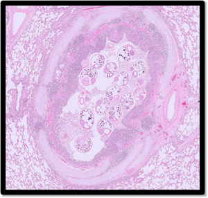
  • Lung samples were received from a seven-month-old pig that had died suddenly and, on postmortem examination, a large volume of pleural fluid was found around the lungs. There were clinical concerns over the possibility of Glasser’s disease or other causes of septicaemia. On histopathology, adult nematodes were found within the bronchi, consistent with verminous bronchitis following infection with Metastrongylus lungworm species (Figure 5). Infection with this species may or may not be associated with clinical signs and, as there were minimal inflammatory changes present, was probably not of great significance to this individual. Oedema was also present within septal and subpleural tissue and to a lesser extent the pulmonary parenchyma. There was no evidence of fibrinous pleuritis to suggest Glasser’s disease. Pulmonary oedema may occur secondary to systemic inflammatory response although no clear underlying cause for it was evident in the sections examined and other conditions such as heart failure leading to pulmonary oedema could not be excluded.

Figure 5: Adult lungworms in the airway of a lung of a pig.

  •  

Deficiencies and toxicities

  • Bloods were submitted from two Jersey cows from a herd which were having issues with retained placentas and metritis following Both cows had raised BHB (2.32mmol/l, 5.26 mmol/l, RI= 0.00-1.2mmol/l) and NEFA levels (1980 µmol/l,1240 µmol/l, RI= 0-600µmol/l) indicating ketosis and evidence of excessive fat mobilisation. NEFA concentrations of more than 1000 µmol/l can be associated with fatty liver and increase the risk of milk fever, ketosis, infertility and displaced abomasum. Fatty liver syndrome arises from cows getting too fat during late pregnancy and is a very important cause of appetite suppression.
  • Ragwort toxicity was suspected in a 14-month-old bovine which had shown signs of reduced mentation, head pressing, blindness and tenesmus and progression to seizures and death. It was the second such case to occur in the group. Histopathology identified a severe, chronic, diffuse, hepatopathy with biliary hyperplasia, portal bridging fibrosis, and rare hepatocyte megalocytosis. The distribution and nature of the lesions were considered most consistent with a chronic toxic insult and in the UK this is most frequently associated with ragwort poisoning. Mycotoxins were suggested as another possible cause. The blood lead measurement was a background levels indicating there was no exposure to a lead source.
  • Hypocalcaemia was identified as the likely cause of sudden death in a freshly calved dairy cow by measurement of calcium in aqueous Calcium levels of <1mmol/l in aqueous or vitreous humour are suggestive of terminal hypocalcaemia.
  • Low Vitamin B12 levels indicative of cobalt deficiency was identified in numerous cases in July and August. In one case seven of eight homebred mule yearlings which were failing to thrive had low B12 level (RI >221pmol/L). The plasma inorganic iodine level in a pooled sample from six of the sheep had a low PII level of 36µg/L indicating insufficient recent intake of iodine (PII levels of 60-150µg/l indicates marginal iodine intake in animals contributing to the pooled sample and levels >150µg/l indicate adequate intake). Low PII in a pooled sample indicates there may be an increase of risk of clinical disease and production issues related to iodine deficiency in that group.
  • There were multiple cases of low copper results suggestive of copper deficiency in July and August. Very low copper levels (1.1 and 6 µmol/l: RI 9 – 25) were reported in two goat kids that had gone off their legs at three months of age, consistent with delayed swayback.
  • A two-month-old Aberdeen Angus cross bull calf showed signs of stiffness in the hindlimbs which then progressed to involve the forelimbs. Testing of the blood samples submitted identified evidence of hypovitaminosis E with a Vitamin E level of 7mg/l (RI 1.3-7.8mg/l) and hypocupraemia with a copper level of 3.3µmol/l (RI 9.0 -19.0µmol/l)
  • Low GSH-Px levels indicative of selenium deficiency were identified in three of four tested dairy heifers (the fourth had a GSH-Px result just above the cut off value of 30U/ml RBC) from a group which were failing to conceive despite appearing clinically normal. Selenium deficiency was also suspected in a yearling animal with a stiff gait, which had a serum selenium level of 639nmol/l, just below the reference value of 640-1020nmol/l.
  • A low serum zinc level of 2µmol/l (RI 12.2-18.2µmol/l) was measured in a post weaned Jersey calf with signs of hyperkeratosis. Low serum zinc levels were also reported in at least six goats on different holdings, four of which were pygmy goats with a history of skin disease.
  •  
  •  

Follow us on Facebook

https://www.facebook.com/Axiomfarmvetlab/

This newsletter is sent by e-mail to each vet practice but if you would like a copy sent to your individual e-mail account please contact us at dsfarm@axiomvetlab.co.uk and we can add you to our circulation list.

Related Posts