Farm August Newsletter 2026

Dedicated farm line – 01626 357776

Follow us on Facebook

https://www.facebook.com/Axiomfarmvetlab/

This newsletter is sent by e-mail to each vet practice but if you would like a copy sent to your individual e-mail account please contact us at dsfarm@axiomvetlab.co.uk and we can add you to our circulation list.

Would you like to send the Axiom farm vets photos of PMEs to allow us to assist with sample selection and diagnosis? Please send photos by WhatsApp to 07944 649 222.

Note that we may wish to include photos sent to this number for the interesting cases section of this newsletter. If you do not wish your photo to be used, please let us know at the time of sending.

Email us with your additional test requests

You may find this is a more efficient way of making requests than phoning the farm team, saving you time in your busy day. Our farm team also find it a more efficient way of dealing with your requests.
The email address for test requests is: dsfarm@axiomvetlab.co.uk 

BTV testing update and testing guidance

Official figures just released by the APHA indicate that there have now been over almost 500 cases of confirmed BTV-3 infection in England (as of 21/08/26), with 15 cases also confirmed in Wales and 6 in Scotland. In England, most cases have occurred in the southwest with over 300 confirmed cases in Devon alone and more than 150 cases in both Cornwall and Somerset. Many more cases than this have been reported and there is a 10-day lag in the figures due to the testing time required at Pirbright.

Livestock, particularly sheep, are getting severe illness, with clinical signs including facial swelling, conjunctivitis, drooling, oral lesions, scour, lameness, abortions, teat lesions and pyrexia (40-41+ oC). Pirbright have reported that the long spell of hot, dry weather has sped up viral replication in midges. They have also calculated that infected midges need to be exposed to a total of 50.7 degree-days for the replication in midges to be completed (data for BTV-8) and the Met Office’s Atmospheric Dispersion and Air Quality (ADAQ) team have produced a map that illustrates that this threshold has been passed in nearly all of England, including the north, the whole of Wales and large parts of southern Scotland and Northern Ireland.

BTV made it to Cumbria last year and there have already been a few cases in northern England this year. There is a high risk that BTV-related disease will be experienced by many sheep and cattle farmers across the country. There is still time to prepare and vaccinate for what is probably coming to your clients’ farms. Stocks of vaccine in the UK are currently very low. Some vaccine is being imported from Europe under licence but there will be a delay before this arrives. It would be advisable to encourage farmers to place orders, so the manufacturers have an idea of demand, although this is unlikely to change vaccine availability in the short term.

Please note that, due to SAPO regulations, we cannot accept samples from animals showing signs suspicious of Bluetongue infection, being tested for Bluetongue or that have recently tested positive for Bluetongue. If such samples are received, they will have to be destroyed. We can accept samples from herds or flocks which are currently undergoing testing for BTV or are showing signs consistent with BTV infection provided there is no suspicion of bluetongue virus infection in the individual being tested. We are happy to discuss cases over the phone before submission of samples. Clinical suspicion of BTV infection must be reported to APHA.  For the latest information and updates on BTV in the UK please visit the following link: Bluetongue: latest situation GOV.UK

BVD Cymru Eradication Programme

Compulsory legislation for BVD eradication in Wales has now been implemented. The rules require all cattle keepers to complete annual BVD screens and movement restrictions from non-tested or infected herds will be enforced. Furthermore, BVD test results for both screening and diagnostic submissions must be uploaded to the BVD Cymru database. Practices are therefore reminded that for all submissions from Welsh farms requesting BVD testing (apart from bulk milk testing), even as part of a testing package, will also require a BVD Cymru submission form to be submitted along with the full UK ear tag number of the animal(s) being tested.

Axiom Farm Factsheets

Did you know there are lots of farm animal factsheets available on the Axiom Veterinary Laboratories website? These can be downloaded and used as handy guides when making decisions on sampling and testing. The factsheets can be accessed using the link below or scan the QR code in the following article on farm histopathology: Farm Animal Fact Sheets Axiom Veterinary Laboratories

Farm histopathology

We offer farm histopathology on postmortem samples, and on biopsy samples from live animals under the code FPM01. Up to three different tissues can be processed under this code and extra tissues can be processed on request. Where different lesions/ stages of disease are seen in one organ (e.g. pneumonic lungs) multiple small sections can be submitted as one tissue type and this will not incur additional costs. For best results only perform histopathology on tissues from fresh carcases and ensure samples are transferred into fixative as soon as possible after sampling. Samples which have been submitted as fresh tissues cannot be processed for histopathology as there is likely to be severe autolysis by the time they reach the laboratory.

Formalin containing histopathology pots can be ordered with other consumables through the website. Scan the QR code below to access our ‘How to get the most out of your large animal histopathology service’ factsheet. Further tips on sampling for histopathology can be found in the ‘Histopathology: points to consider’ and ‘Sampling for respiratory disease’ factsheets.

Feedback on our services

As part of our continuing effort to help improve our services to clients within the Laboratory Division we have put together a short survey which will be sitting live permanently for anyone to complete whenever they have a few minutes spare. This can be filled in by anyone who uses any of our services and we encourage feedback to help us understand what we are doing well and where we need to make improvements. The QR code and link to the survey will remain live as a continuous tool to enable us to always gather feedback. If anyone has any questions about this, they are welcome to contact either of the Quality Managers at the Laboratories, Claire Richardson for Axiom Veterinary Laboratories and Susan Reeve for Finn Pathologists. Thank you in advance for helping us to improve our services.

https://www.surveymonkey.com/r/Laboratory_Satisfaction_Survey

Farm Post -Mortem CPD: Book now!

Interesting cases in June

Cattle
Abortions/mastitis

  • BVD virus was detected by PCR in foetal spleen tissue from one case. The 600-head flying herd had experienced four premature calvings or stillbirths in the previous week, and multiple cases of pyrexia had been identified in the heifer group, raising the suspicion of active BVD infection.
  • Pseudomonas aeruginosa was isolated in cultures of foetal stomach contents in one case of abortion. Bovine abortion due to aeruginosa is rare and usually sporadic with the bacterium acting as an opportunistic pathogen. Ascending infection from the lower genital tract or infection via the maternal bloodstream are both possible.
  • Streptococcus pleuranimalium was isolated from the aborted foetus of a British blue cross This opportunist pathogen has been associated with several different conditions in domestic ruminants (and humans), including abortions.
  • Salmonella Dublin was isolated in two abortion cases both in dairy cattle in the seventh month of
  • Coxiella burnetii, the causative agent in Q fever, was detected by PCR testing of a vaginal swab in an abortion case at five months The abortion occurred at pasture, and the foetus and afterbirth could not be found. Detection of the agent in vaginal swabs indicates that C. burnetii was being shed from the reproductive tract but cannot confirm it as the cause of abortion. The dam was reported to be clinically well.
  • Listeria monocytogenes was isolated from milk from a cow with a persistent high cell Listeria monocytogenes is a relatively rare cause of mild to severe suppurative mastitis in cattle.
  • Pasteurella multocida was isolated from mastitic milk in one P. multocida is a rare cause of mastitis in cattle but can be severe with a high risk of endotoxaemia and often a poor response to antimicrobial treatment.
  •  

Respiratory disease

  • IBR was detected by PCR in at least five cases this Affected animals showed typical signs of pyrexia, nasal and/or ocular discharge, and respiratory distress.
  • Severe lung consolidation and pleurisy with interstitial oedema were seen during postmortem examination following the sudden death of a 54-day-old Aberdeen Angus suckler Samples of lung tissue submitted tested positive for Mannheimia haemolytica by PCR. The findings were considered consistent with a case of acute M. haemolytica pneumonia/ septicaemia. Testing of liver tissue identified marginally low selenium levels, raising the possibility that low selenium status could have been a contributing factor in the case by causing increased susceptibility to secondary infection.
  • Dictyocaulus viviparus (lungworm) larvae were detected by PCR testing of a pooled faecal sample from three 22-month-old Monbeliard heifers with signs suspicious of lungworm infection post The heifers had received a lungworm vaccine. The significance of the finding of lungworm was uncertain as although vaccination is expected to prevent clinical signs it does not prevent small numbers of lungworm from establishing. It was advised that if the signs seen were consistent with lungworm infection that the vaccination protocol should be reviewed (e.g storage, dose, techniques, timings between doses and between the second dose and turnout). Vaccine failure is classified as an adverse event and if suspected should be reported to the Veterinary Medicines Directorate (VMD). If any anthelmintics had been given these could have reduced the efficacy of the vaccine. It was also suggested that any factors affecting the immune system could be worth investigating e.g BVD status, copper, selenium, iodine and vitamins A/E. Due to the dry summer there have been relatively few lungworm cases diagnosed in the laboratory but this could quickly change if wetter weather arrives in September.

Gastrointestinal disease

  • An eight-month-old Holstein heifer showed signs of scour. No other animals in the group were Ivermectin treatment (route of administration not stated in history) had been given two weeks previously but the animal had not improved. A faecal worm egg count was performed and there was a strongyle egg count of 700 epg indicating a heavy gastrointestinal nematode burden. No fluke eggs were seen on microscopy and Salmonella cultures were negative. The high worm egg count in the recently treated animal was suggestive of anthelmintic treatment failure. Possible reasons for treatment failure include underdosing, technique used, failure to calibrate equipment or equipment malfunction, incorrect storage or use of medicine past the expiry date. If these failures were ruled out, the next step would be to consider performing a faecal egg count reduction test (FECRT) to investigate ivermectin resistance.
  • Cryptosporidiosis caused watery scour and depression in ten-day-old dairy calves on one
  • Salmonella Montevideo was cultured from a faecal sample from dairy calves which were scouring. Bovine coronavirus antigen was also detected in the sample and was likely to be significant in the clinical presentation in the calves. The submitting vet was also aware of Salmonella infections in the adult herd in this case.
  • Salmonella Typhimurium was isolated from a pooled faecal sample from three adult Holstein-Friesian cattle with pyrexia and profuse bloody scour. Liver fluke and rumen fluke eggs were also observed on faecal
  • A two-year-old hired bull which had arrived at the current herd in March and had been at pasture serving cows showed signs of weight loss which had become more significant in the previous two weeks. At the time of examination, the bull was scouring and was not interested in eating concentrate feed. Gut sounds were reduced and the animal appeared thin. Johne’s PCR was positive, consistent with a diagnosis of Johne’s disease with the test Ct value of 23 indicating heavy shedding of MAP bacteria. If the bull had been tested prearrival , or in quarantine on arrival, he would likely have been identified on serology as being a risk to the herd.

Skin and eye disease

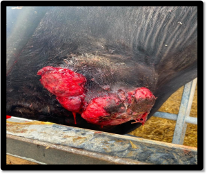
  • An Actinobacillus was isolated from a swab of a mass on the shoulder of a yearling bullock. The lesion was described as slow growing and was discharging purulent material (Figure 1). The animal was otherwise well in itself. MALDI ToF analysis is unable to reliably differentiate between species of the genus Actinobacillus, but in this case, it was considered likely to be Actinobacillus lignieresii, the pathogen responsible for cutaneous actinobacillosis. Cutaneous actinobacillosis was also diagnosed in another case in a limousin cross heifer, which was also showing signs of a slow growing mass on the shoulder. Biopsy samples from the mass were submitted for histopathology which identified a fibrosing and granulomatous dermatitis/ cellulitis with appearance consistent with a case of cutaneous actinobacillosis.

Figure 1: Lesion on the shoulder of a yearling bullock due to suspected cutaneous actinobacillosis. (Photo : Rebecca Edwards, Carisbrooke vets)

  • Pseudomonas aeruginosa was isolated in swabs from the eyes of yearling calves with signs of conjunctivitis and a poor response to parenteral oxytetracycline. aeruginosa can act as an opportunistic pathogen in eye disease and so the finding was considered of likely significance in this case. As is common with Pseudomonas species, which have innate resistance to many antibiotics, the isolate in this case showed in vitro resistance to multiple antimicrobials including oxytetracycline.

Systemic and miscellaneous disease

  • Mucosal disease following persistent BVD infection was suspected to be the cause of severe haemorrhagic diarrhoea and pyrexia in a yearling heifer at A serum sample tested positive for BVD antigen in the ELISA with a high positive result.
  • BVD virus was detected by PCR in a neonatal calf which was born blind and had other neurological deficits. The low Ct value of 26 indicated a high level of virus The animal was suspicious of being a BVD PI, though a second test (usually BVD antigen ELISA once the calf reaches one month of age) could be used to definitively confirm BVD PI status.
  • Babesiosis was diagnosed on examination of a blood smear from a cow in Northumberland which was dull and anaemic. There was a recent history of babesiosis cases on neighbouring farms.
  • Three adult cows at grass showed signs of lethargy, mild tachypnoea and mild hock There was a history on farm of tickborne disease. Of the three cows tested one was positive for Anaplasma phagocytophilum by PCR testing of EDTA blood samples confirming Tickborne fever (TBF) in that animal. On another farm, of a group of 38 ten-month-old British blue cattle, approximately six had signs of pyrexia and high heart rates. Otherwise, the clinical examination was unremarkable, but ticks were seen on the animals. Two of three tested animals were positive for antibodies to Anaplasma phagocytophilum.
  • An adult Holstein-Friesian dairy cow showed signs of being off-colour post calving. Blood samples were submitted and on biochemistry GLDH and GGT were significantly raised indicating evidence of hepatocellular injury and hepatic cholestasis. NEFA’s were severely raised at a level of 2000µmol/l (RI = 0-600µmol/L) indicating excessive mobilisation of fat stores. Fatty liver disease was suspected to be the cause of the clinical signs and raised liver enzymes in this case.
  • Formalin fixed samples of brain were submitted to investigate the death of a three-month-old South Devon suckler calf following acute onset neurological signs of lateral recumbency and paddling. The owner was concerned about potential for tick borne disease in the herd. Histopathology revealed a multifocal acute, necrotising encephalitis with a fibrinohaemorrhagic and suppurative meningitis. An infectious, haemotogenous aetiology was considered most likely and suggested potential causes included Escherichia coli, Streptococcus spp., Staphylococcus spp. and Salmonella spp.
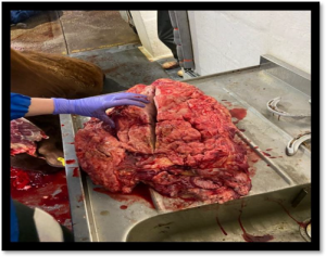
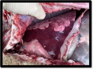
  • Postmortem material was received from a fourteen-month-old Sussex steer which was euthanased after presenting three weeks earlier with an acute and rapidly enlarging swelling in the ventral (Figure 2). The mass was identified on histopathology as thymic tissue infiltrated by an unencapsulated, densely cellular proliferation of neoplastic round cells. A lymphoid rich thymoma or thymic lymphoma were the main differentials, and thymic lymphoma was considered the most likely diagnosis. Thymic lymphomas in cattle are usually T cell lymphomas, affect animals 6-24month of age and are usually sporadic.

Figure 2: Postmortem appearance of a suspected thymic lymphoma removed from ventral neck of a fourteen-month-old steer (Photo: Abigail Buck, Fairfield farm vets)

Sheep and goats

Abortions, reproductive disease, mastitis

  • A haemorrhagic mass found in the vagina of a five-year-old female Toggenberg goat was found to be a haemorrhagic and inflamed vaginal polyp on histopathology. These are non-neoplastic, benign lesions, can be single or multiple and often arise from the ventral floor of the vagina. It was difficult to confirm complete excision from the sections examined and monitoring for recurrence was advised.
  • Several cases of Staphylococcus aureus and Mannheimia haemolytica mastitis in ewes were reported this month, these being the most common causes of severe mastitis in In one flock, 100 ewes had been lambed, of which 20 had developed gangrenous mastitis. Two milk samples were submitted, and S. aureus was isolated from one and M. haemolytica from the other.
  •  

Respiratory disease

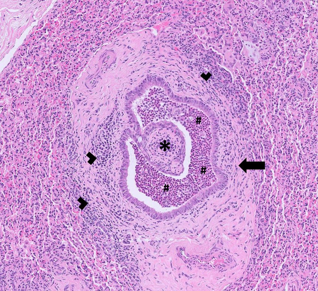
  • Formalin fixed samples of lung from a lamb were submitted to investigate the cause of respiratory signs in a flock in which Mycoplasma involvement was suspected (Figure 3) Histopathology detected severe, suppurative bronchointerstitial pneumonia, consistent with a severe aerogenous bacterial insult likely secondary to an infection with Mycoplasma ovipneumoniae, with differentials including Bibersteinia trehalosi, Pasteurella multocida and Mannheimia haemolytica (Figure 4) Concurrent or prior viral infection (e.g. PI3v and RSV) also could not be definitively excluded. M. ovipneumoniae is recognised as a primary agent in atypical (chronic, non-progressive) pneumonia, usually associated with mild or subclinical respiratory disease in lambs during their first year of life. This condition is often multifactorial in nature and is influenced by a range of management and environmental factors, including mixing of animals, high stocking density and suboptimal air quality. Although such lesions are more commonly encountered during winter months, when predisposing conditions are prevalent, similar changes may also be identified in grazing animals. In cases with persistent coughing rectal prolapse can occur.


Figure 3: Atypical pneumonia in a lamb (Photo: Naomi Church, Hill and Phelan vets)
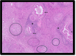
Figure 4: Airway epithelium hyperplasia (arrows), airway distortion (circles), lymphoplasmacytic infiltrate (*), and alveoli filled with neutrophils (- – -) in the lung of a lamb with atypical pneumonia.

  • One case of OPA diagnosed on histopathology this month was in a four-year-old ewe, one of six to have died acutely in the previous four days. The ewes had been bought in, their vaccination status was unknown, they had recently received anthelmintic and there was no known access to toxins, the field having been grazed by sheep for some years. On PME, the lungs looked abnormal and fibrinous pleural fluid was present. Changes were typical of OPA on histopathology, with evidence of a secondary bacterial pneumonia, consistent with the isolation of Mannheimia haemolytica on culture, and which would have accounted for the sudden death in the ewe.
  • In a sheep flock of 100 animals, five lambs had died in a week, affected animals showing respiratory signs 24 hours prior to The lambs received two doses of Ovivac P, the second dose three weeks previously. PME of one lamb found marked consolidation of the lungs with some rubbery areas present. The histological findings were consistent with chronic bronchointerstitial pneumonia with evidence of a bacterial bronchopneumonia component. However, there were also atypically large Type II pneumocytes and abundant foamy macrophages, suggesting that the lamb was showing early signs of developing OPA. There was a history of OPA in the flock.
  • Muellerius capillaris was detected on Baermann’s testing of a faeces sample from a female Toggenberg M. capillaris is more pathogenic in goats than sheep and clinical signs range from a mild cough to a severe chronic cough and dyspnoea. Protostrongylus rufescens was detected on Baermann’s testing of a faeces sample from a heavily pregnant ewe; this species can cause respiratory signs in sheep if heavy burdens are present.
  • Further cases of Maedi visna infection were picked up on serology this month. Including a ewe in poor body condition and with chronic respiratory disease.

Gastrointestinal disease

  • A group of lambs of mixed sizes out grazing with ewes were scouring and struggling to put on condition, with some dying acutely. The lambs had been treated for coccidiosis with limited success. A coccidial oocyst count of > 50,000 opg on a pooled faeces sample from 10 lambs contained 10% Eimeria ovinoidalis and 5% crandallis, the two pathogenic Eimeria species in sheep, confirming continued issues with coccidiosis.
  • Several cases of parasitic gastroenteritis were diagnosed this month in sheep and goats, with clinical signs including scour and ill In one flock, where ewes and lambs were recumbent, with pale mucous membranes and dying, a pooled sample from a mix of ages of sheep was received. Of a strongyle egg count of 2,700 epg, 18% were identified as being Haemonchus sp. by the Ovacyte method and liver fluke eggs were also seen, indicating concurrent fasciolosis. In another flock, Nematodirus battus eggs were detected in the faces of a scouring lamb, which also had a high coccidial oocyst count although no oocyst speciation was carried out. Further cases of fasciolosis were also reported in other flocks this month.
  • Among the cases of Johne’s disease was a two-year-old female goat with a history of chronic weight loss and some scour that The goat had pale mucous membranes and originally had been diagnosed with haemonchosis (faecal strongyle egg count of 2393 epg, with 42% Haemonchus). However, the weight loss continued and screening for Johne’s was therefore carried out.
  • Salmonella 61:k:1,5,7 was isolated from a faeces sample from a scouring two-year-old This is the most common serovar isolated from sheep and its clinical significance is unclear as it may be isolated from healthy as well as scouring sheep.

Skin and eye disease

  • Orf was confirmed on PCR testing of scab material from a two-year-old
  • Skin scabs and a swab were received from a seven-year-old male goat with ongoing scabs around the nostrils which, after seven months, had progressed on to the top There were also some scabby flakes on the body. Blood zinc was within normal range last year and there was no improvement after treatment with Imaverol. Staphylococcus aureus was isolated from the skin swab. Staphylococcal dermatitis infection is common in goats, although more typically found on udder, teats and groin.
  • A skin biopsy was submitted from a nine-year-old ram to investigate a large mass on the thorax which was infiltrating the underlying musculature. Histopathology was consistent with melanoma. Melanocytic neoplasms are considered uncommon in sheep, with malignant melanoma representing a rare diagnosis in this species. Reported cases indicate that these tumours most frequently arise in the skin and mucocutaneous junctions but may also occur in visceral locations, sometimes without identification of a primary lesion.
  • Lice were submitted from goats in a mixed enclosure with ponies on a farm The infestations had started in the goats and multiple treatments with ivermectin had been carried out. The lice were identified as the biting louse, Bovicola ovis, which, although more commonly found in sheep, can persist and reproduce on goats which act as a possible reservoir of infection.

Systemic and miscellaneous disease

  • Clostridium perfringens epsilon toxin was detected in the ileal contents of an unvaccinated six-week-old lamb that died suddenly, potentially consistent with pulpy kidney, although brain histopathology is required to confirm diagnosis.
  • Seroconversion to CAE was detected in two of ten Golden Guernsey goats screened before entry to an agricultural
  • Following the death of five sheep in a flock in Devon with no premonitory clinical signs, an EDTA blood sample was received from a pyrexic shearling ewe from which Anaplasma phagocytophilum was detected, confirming Tick Borne Fever.
  • Postmortem tissues were received from a ewe exhibiting neurological signs and histopathology of brain revealed a severe, necrotising polioencephalopathy most suggestive of cerebrocorticonecrosis (CCN), caused by thiamine deficiency or sulphate The former is commonly associated with recent changes in diet resulting in rumen dysbiosis and the production of thiaminase with subsequent vitamin B1 deficiency. Sulphate toxicity is associated with excess intake either through concentrate food and/or water.
  • CLA was confirmed on culture of swabs from abscesses in a goat and a sheep and seroconversion to CLA was reported in several In one flock, all rams were screened after two had tested positive the previous week. The rams had all been together four weeks previously and sheared at the same time. Six of the 31 rams were seropositive.
  • Klebsiella aerogenes was isolated following enrichment culture of a swab from an abscess in a sheep. Klebsiella species are associated with wound infections, mastitis and abscesses in farm animals and aerogenes, formerly known as Enterobacter aerogenes, is a rare opportunistic pathogen in the species.
  • Trueperella pyogenes was isolated from a swab taken from a purulent infection in the ear of a
  • Post-mortem tissues were received from a two-month-old lamb which died following a four-day history of lethargy, inappetence and respiratory distress. The lamb was unvaccinated and failed to respond to NSAIDs, antibiotics and fluids. PME found ulcers on the muzzle, consolidation of cranial lung lobes and pale areas (infarcts) in the epicardium. The case had been notified to APHA and BTV testing was negative. On histopathology, there was severe, extensive myocardial fibrosis with mineralisation and ongoing myofibre degeneration and necrosis most likely consistent with White Muscle Disease. There was also severe, necrosuppurative, lymphohistiocytic cheilitis, folliculitis and vasculitis with intralesional bacteria associated with the muzzle ulcers which was suspected to be secondary to the heart lesions as vitamin E / selenium deficiency can damage blood vessels. Finally, there was severe, subacute suppurative bronchopneumonia with intralesional plant fragments and debris which was likely to be a secondary/ terminal bacterial bronchopneumonia following terminal aspiration.
  • An 11-year-old female pygmy goat lost weight and became ataxic, with head pressing. The animal’s condition deteriorated and it was euthanased. PME only found watery abomasal and intestinal contents and heat stroke was suspected as the condition started during a period of extremely hot weather. Blood samples taken before euthanasia revealed an azotaemia with a discordantly high urea and elevated liver enzymes. On histopathology, there was evidence of multifocal splenic necrosis/infarction, multifocal tubular nephrosis and multifocal acute hepatitis with individual cell necrosis. Random individual cell necrosis in the liver suggested a reactive hepatitis secondary to a systemic insult and explained the elevation in liver enzymes. There was also evidence of hepatocellular swelling in intact hepatocytes suggesting degrees of injury ranging from sublethal to The spleen is a target organ for damage in heatstroke and while splenic inflammation/infarcts may occur secondary to septicaemic or toxaemic processes, it is a more unusual finding, therefore the suspicion that the episode of illness was triggered by extreme hot weather was supported by this finding. The tubular nephrosis also correlated with the biochemical findings and suggested multiorgan failure secondary to a heat stress or stroke induced problem in this goat. Liver copper analysis also revealed that the goat was also suffering from copper deficiency.


Camelids

  • A 17-year-old female alpaca became recumbent, was hypothermic and had pale mucous A count of 100 opg Eimeria macusaniensis was detected in a faeces sample. Although this is a low count, with E. macusaniensis having a prepatent period that can exceed 30 days, this may still have been significant. E. macusaniensis is the most pathogenic Eimeria species in South American camelids, causing clinical coccidiosis in adults as well as crias.
  • Fasciolosis was diagnosed on liver fluke coproantigen ELISA testing in a three-year-old alpaca that was losing weight and was in very poor body condition.
  • Johne’s disease was diagnosed on PCR testing of faeces from a five-year-old male alpaca with watery diarrhoea for two
  • A 12-year-old neutered male alpaca was straining to urinate and had a poor flow rate. Solid material was visible in the turbid urine sample submitted and urinalysis revealed a small number of triple phosphate (struvite) crystals and a large amount of amorphous debris. As with sheep and cattle, neutered males are at increased risk of obstruction because of a narrower urethra and the long urethra in male camelids and low urine output in these animals potentially further increases the risk of obstruction. Cebra (2014) found no uroliths in about half the cases of urethral obstruction in his clinic and suggested that obstruction may be more a consequence of prostatitis or cystitis. (Ref: Cebra, C. (2014). Disorders of the urinary system. In Llama and Alpaca Care (pp. 464-476). WB Saunders.)

Other species

  • Fasciolosis was diagnosed in a reindeer on liver fluke coproantigen ELISA testing. No clinical history was
  • Salmonella Senftenberg was isolated from a yolk swab taken from a pheasant flock with enteritis throughout the group, issues with dirty eggs and some yolk sac infections. This serotype is one that the APHA does isolate occasionally in pheasants as well as other poultry species. In a second case of yolk sac infection in one-week-old pheasants, Enterococcus faecium and coli were isolated, both of which were likely to be significant opportunist pathogens. An insufficiently humid hatching environment has been associated with a high incidence of yolk sac infection.
  •  

Deficiencies and toxicities

  • A poor calving season was reported in one beef suckler herd with adults in poor body condition and two down cows after calving which failed to recover with The cows had been moved frequently during the calving period due to lack of available forage. Despite the poor condition of the cows, the calves appeared satisfactory. Six cows were sampled for metabolic profiles and trace element testing. Several cows were identified as hypoalbuminaemic and all had high urea levels. In metabolic profiles multiple animals with low albumin and high urea suggests there is an overall shortage of metabolisable protein due to insufficient rumen non degradable protein, or due to an excess of rumen degradable protein that cannot be utilised due to a lack of fermentable metabolisable energy (energy available to rumen microbes). All the cows had evidence of hypocupraemia. Three of the six had copper levels well below the threshold of 6µmol/l where clinical copper responsive disease is likely to occur. All but one had low GSH-Px levels consistent with selenium deficiency. The pooled plasma inorganic iodine level for the group was low, indicating low recent dietary iodine intake of the group and an increased risk of disease due to clinical iodine deficiency.
  • On one farm three dairy calves under ten days of age were showing signs of blindness and had evidence of cataracts on clinical examination but appeared otherwise The dry cows had recently been turned out to grass and had access to mineral buckets but were not being given any concentrate feed. Two of the calves were tested and both had low Vitamin A levels at 140µg/l and 203µg/l (RI = 249-500µg/l). One calf was tested for immunoglobulin levels in the ZST test and had a marginal ZST level of 19 ZST unit indicating that colostral intake may have been suboptimal. This finding could be significant as colostrum is an important source of Vitamin A for the newborn calf. Therefore, Vitamin A deficiency in pregnancy could not be confirmed or ruled out as the cause of the eye defects in the calves. BVD vaccination was not used in the herd. BVD testing indicated that the calves had neither seroconverted to BVD or received maternal antibodies to BVD via colostrum. One of the three calves was tested for BVD virus by PCR on serum and there was no evidence of BVD viraemia. These results suggested that BVD infection was unlikely to be involved in the developmental eye abnormalities in this case. In a different herd four calves from a spring block calving herd were showing signs of poor growth and all had vitamin A levels below the reference interval.
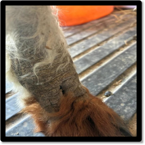
  • Skin biopsy samples from a two-year-old alpaca with a non-pruritic hyperkeratotic skin disorder were submitted for histopathology (figure 5). No ectoparasites had been seen on scrapes despite multiple skin scrapes being A blood sample taken previously had been tested for zinc levels which were found to be below the reference interval at 1.8µmol/l (RI=3.0-14.6µmol/l). Examination of the biopsy samples revealed features consistent with a keratinising disorder. Zinc responsive dermatosis in alpaca may be due to true zinc deficiency or a keratinising disorder that is responsive to high doses of zinc supplementation. This has also been termed idiopathic hyperkeratosis. Lesions in affected animals consist of thickened skin with a tightly adherent crust often on the ventral abdomen, axilla, medial thigh and inguinal area but also on the face, pinnae and neck in some cases. Dark fleeced animals and young females are more frequently affected. There were at least two other cases of low blood zinc levels on alpaca this month; in one of these animals the larval stages of Chorioptes sp. mites were also observed in skin scrapes.
  • There were several cases of lead toxicity recorded in cattle this Lead poisoning cases tend to increase during the summer months when the chance of exposure to a lead source (e.g a discarded car battery) is increased. On one farm there were three sudden deaths of 14-month-old cattle, and a further animal was found down with bilateral blindness, twitching and watery diarrhoea. No significant lesions were seen on postmortem. Both the blood lead results in the live animal and the kidney tissue level from the postmortem samples were consistent with a diagnosis of lead toxicity.
  • Skin biopsy samples from a two-year-old alpaca with a non-pruritic hyperkeratotic skin disorder were submitted for histopathology (figure 5). No ectoparasites had been seen on scrapes despite multiple skin scrapes being A blood sample taken previously had been tested for zinc levels which were found to be below the reference interval at 1.8µmol/l (RI=3.0-14.6µmol/l). Examination of the biopsy samples revealed features consistent with a keratinising disorder. Zinc responsive dermatosis in alpaca may be due to true zinc deficiency or a keratinising disorder that is responsive to high doses of zinc supplementation. This has also been termed idiopathic hyperkeratosis. Lesions in affected animals consist of thickened skin with a tightly adherent crust often on the ventral abdomen, axilla, medial thigh and inguinal area but also on the face, pinnae and neck in some cases. Dark fleeced animals and young females are more frequently affected. There were at least two other cases of low blood zinc levels on alpaca this month; in one of these animals the larval stages of Chorioptes sp. mites were also observed in skin scrapes.

Figure 5: crusting and scaling of the skin of the lower limbs in an alpaca with suspected zinc responsive dermatosis. Similar changes with less severe thickening were seen on the dorsum of the neck, in the axilla, inguinal and perineal areas. (Photo: Jenny Schmidt, Torch farm vets)
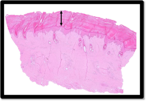
Figure 6: Marked orthokeratotic hyperkeratosis (arrow) of the skin in an alpaca suggesting a zinc responsive dermatosis

  • A low zinc level in blood of 8µmol/l (RI > 9.9µmol/l) was measured in a female pygmy goat with crusting and scaling of the skin around the tail base. There were at least five other cases of low blood zinc levels in goats this month, three of which were also pygmy goats.
  •  

Follow us on Facebook

https://www.facebook.com/Axiomfarmvetlab/

This newsletter is sent by e-mail to each vet practice but if you would like a copy sent to your individual e-mail account please contact us at dsfarm@axiomvetlab.co.uk and we can add you to our circulation list.

Related Posts