Farm July Newsletter 2026

Dedicated farm line – 01626 357776

Follow us on Facebook

https://www.facebook.com/Axiomfarmvetlab/

This newsletter is sent by e-mail to each vet practice but if you would like a copy sent to your individual e-mail account please contact us at dsfarm@axiomvetlab.co.uk and we can add you to our circulation list.

Would you like to send the Axiom farm vets photos of PMEs to allow us to assist with sample selection and diagnosis? Please send photos by WhatsApp to 07944 649222.

Note that we may wish to include photos sent to this number for the interesting cases section of this newsletter. If you do not wish your photo to be used, please let us know at the time of sending.

Email us with your additional test requests

You may find this is a more efficient way of making requests than phoning the farm team, saving you time in your busy day. Our farm team also find it a more efficient way of dealing with your requests.
The email address for test requests is: dsfarm@axiomvetlab.co.uk 

BVD Cymru Eradication Programme

Compulsory legislation for BVD eradication in Wales has now been implemented. The rules require all cattle keepers to complete annual BVD screens and movement restrictions from non-tested or infected herds will be enforced. Furthermore, BVD test results for both screening and diagnostic submissions must be uploaded to the BVD Cymru database. Practices are reminded therefore that for all submissions from Welsh farms requesting BVD testing (apart from bulk milk testing), even as part of a testing package, will also require a Welsh BVD submission form to be submitted along with the full UK ear tag number of the animal(s) being tested.

BTV testing guidance

We wish to remind vets that as a commercial laboratory we are unable to offer any Bluetongue testing either on blood samples or abortion material. If we receive samples from cases in which Bluetongue is suspected, we will be required to safely dispose of the samples without further testing. We are always happy to discuss cases over the phone before submission of samples. Clinical suspicion of BTV infection must be reported to APHA.  Further information on investigating poor reproductive performance in cattle and sheep during Bluetongue outbreaks can be found at the following linkInvestigating poor reproductive performance in cattle and sheep during

Axiom Farm Factsheets

Did you know there are lots of farm animal factsheets available on the Axiom Veterinary Laboratories website? These can be downloaded and used as handy guides when making decisions on sampling and testing. The factsheets can be accessed using the link below or scan the QR code in the following article on farm histopathology: Farm Animal Fact Sheets Axiom Veterinary Laboratories

Farm histopathology

We offer farm histopathology on postmortem samples, and on biopsy samples from live animals under the code FPM01. Up to three different tissues can be processed under this code and extra tissues can be processed on request. Where different lesions/ stages of disease are seen in one organ (e.g. pneumonic lungs) multiple small sections can be submitted as one tissue type and this will not incur additional costs. For best results only perform histopathology on tissues from fresh carcases and ensure samples are transferred into fixative as soon as possible after sampling. Samples which have been submitted as fresh tissues cannot be processed for histopathology as there is likely to be severe autolysis by the time they reach the laboratory.

Formalin containing histopathology pots can be ordered with other consumables through the website. Scan the QR code below to access our ‘How to get the most out of your large animal histopathology service’ factsheet. Further tips on sampling for histopathology can be found in the ‘Histopathology: points to consider’ and ‘Sampling for respiratory disease’ factsheets.

Interesting cases in May

Cattle
Abortions/mastitis

  • Neospora caninum was detected by PCR in foetal tissue in several abortion cases this month. Fresh brain or placenta are the preferred samples for this test, having the highest chance of detection of the parasite in infected calves. Foetal fluid may also be used to test for antibodies to Neospora caninum as the infected foetus will often seroconvert to the pathogen in Seroconversion of the dam to Neospora caninum also raises suspicion that it may be the cause of abortion. In all cases, the diagnosis of abortion due to Neosporosis can only be confirmed by histopathology – particularly of the foetal brain, and heart. Placenta can also be useful for histopathology.
  • There were at least three cases of bovine abortion due to Trueperella pyogenes this T.pyogenes may gain access to the uterus through the bloodstream, originating in lesions such as mastitis or foot abscesses or it may enter through the vagina/cervix. Cases are usually sporadic though occasionally clusters of infections can occur. In one of these abortions, Coxiella burnetii (Q fever) was also detected in placental tissue, but without histopathology the significance of the finding in relation to this abortion was unknown.
  • Filobasidium uniguttulatum was isolated in fungal cultures of a mastitis milk sample. This yeast species has been reported as an albeit rare cause of mastitis. Yeast species often gain entry to the udder because of unhygienic intramammary infusion technique. Sometimes they may act as a true environmental pathogen when present in large numbers in the Treatment with antibiotics is ineffective.
  •  

Respiratory disease

  • Postmortem tissues were received form a one-day-old calf which had received colostrum via stomach tube shortly after birth. The carcase appeared bloated and multifocal patches of consolidated lung tissue were seen grossly which raised a suspicion of a possible aspiration pneumonia. The abomasal mucosa appeared very oedematous with patchy haemorrhagic areas. Histopathology of lung identified a bronchopneumonia with oedema, haemorrhage and foreign material in the airways suspected to be inhaled milk. Examination of abomasal sections found that there was an extensive, marked, acute, fibrinosuppurative abomasitis with intralesional rod-shaped, gram-positive bacteria, consistent with a clostridial abomasitis, possibly associated with tube feeding.
  • Postmortem tissues were received from a two-month-old British-Blue cross suckler calf. Respiratory distress had been observed, and the farmer was concerned about levels of smoke in the shed blowing in from a nearby property. Lesions were seen at postmortem examination that were suspicious of calf diphtheria. Histopathology identified a severe necrosis and inflammation in the soft tissue of the larynx which, along with the clinical history, was considered consistent with necrotic laryngitis (calf diphtheria). Lesions suggestive of subsequent aspiration pneumonia were found in the lung as well as a small, focal area of granulomatous alveolitis. This may also have been due to material inhaled as part of the calf diphtheria but it was not possible to rule out the involvement of smoke inhalation as these changes can be seen with inhalation of environmental allergens/pollutants. Bacteriodes pyogenes was isolated as part of mixed growths in anaerobic cultures of a laryngeal swab and the finding was considered of possible significance as this bacterium is a common secondary invader in damaged tissue. 
  • Histopathology was carried out on lung tissue from a 22-week-old Aberdeen Angus cross bullock which had died In this system calves are brought in at 12-14 weeks of age and sold when they reach 400kg. The group to which this animal belonged had arrived six weeks previously, at which time there had been an outbreak of respiratory disease. Cases had responded well to oxytetracycline, and no further investigation had taken place. Examination of the lung tissue in this case identified extensive, chronic and ongoing changes. There was evidence of longer standing airway damage which may have been caused by a previous pneumotropic viral infection (e.g. BHV 1, RSV, PI 3). There was evidence that this had been followed by a secondary bacterial infection. The finding correlated with results of ancillary testing in which Mannheimia haemolytica was isolated on lung culture also and detected by PCR testing of lung tissue along with Pasteurella multocida and Histophilus somni. In addition, there was a plasmalymphocytic peri-airway infiltrate indicating likely concurrent mycoplasma infection which, considering the negative Mycoplasmopsis (formerly Mycoplasma) bovis PCR result, may have involved Mycoplasmopsis dispar, canis, canadensis and/or alkalescens.
  • Samples were submitted to investigate the cause of death following respiratory signs in a three-month-old beef suckler heifer calf. Bovine coronavirus, Mannheimia haemolytica, Pasteurella multocida and Histophilus somni were all detected by PCR testing. On histopathology more than 70% of the examined tissue was found to be atelectic/collapsed, in association with extensive inflammatory/ necrotic change. Severe bronchointerstitial pneumonia was confirmed with clear histological evidence of bacterial involvement. Alveolar and epithelial changes were also suggestive of pneumotropic viral involvement and earlier infection with e.g. RSV, PI3, could not be ruled out. The coronavirus was considered of doubtful significance. PCR-positive lungs do not necessarily exhibit Bovine Coronavirus-associated pulmonary lesions, and positive results can be the result of luminal debris carried down from the upper respiratory tract. Evidence of underlying selenium deficiency was also found on liver tissue analysis.One-month-old calves which were bought in from market and were being fed on automatic milk machines developed signs of respiratory disease which were pronounced after cold evenings. Coughing, mucopurulent nasal discharge and open-mouthed breathing were observed. Affected calves showed a poor response to tulathromycin injection. Investigations in previous groups had detected evidence of Mycoplasmopsis bovis and vaccination for M.bovis on arrival had been recently initiated. PCR testing of a pooled sample of three nasopharyngeal swabs from this case detected RSV virus indicating active virus infection along with Mycoplasmoposis bovis and Pasteurella multocida. The contribution of the bacterial pathogens to the disease in this case is uncertain as both agents can also be found in the nasopharynx of healthy animals.
  • IBR virus was detected by PCR of lung tissue from an eight-month-old British Blue cross bullock which had been bought in to fatten three months previously. Around 500 animals were on the unit and the submitting vet reported that IBR like signs had been spreading through the They were reported to have received an IBR vaccine on arrival, but further details were not available. In addition to IBR Mycoplasmopsis (formerly Mycoplasma) bovis, Histophilus somni and Pasteurella multocida were also detected and considered significant in this case.
  • One-month-old calves which were bought in from market and were being fed on automatic milk machines developed signs of respiratory disease which were pronounced after cold evenings. Coughing, mucopurulent nasal discharge and open-mouthed breathing were observed. Affected calves showed a poor response to tulathromycin injection. Investigations in previous groups had detected evidence of Mycoplasmopsis bovis and vaccination for bovis on arrival had been recently initiated. PCR testing of a pooled sample of three nasopharyngeal swabs from this case detected RSV virus indicating active virus infection along with Mycoplasmoposis bovis and Pasteurella multocida. The contribution of the bacterial pathogens to the disease in this case is uncertain as both agents can also be found in the nasopharynx of healthy animals.

Gastrointestinal disease

  • Coronavirus was detected in a faecal sample from a two -week-old calf which was scouring and lethargic. Two calves in the same group had died after showing similar Diarrhoea caused by bovine coronavirus typically affects calves from three days to three weeks of age. Outbreaks may be similar or more severe than those caused by rotavirus. Affected calves will have liquid yellow diarrhoea which may contain mucus and blood. As with rotavirus infections, the clinical signs can be exacerbated by concurrent cryptosporidiosis.
  • A nine-year-old suckler cow which calved six weeks previously showed signs of weight loss and Liver fluke eggs were observed on faecal microscopy indicating patent liver fluke infection. Rumen fluke eggs were also seen. The rumen fluke parasite has a similar life cycle to the liver fluke and uses the same intermediate snail host. Mixed infections of liver fluke and rumen fluke are therefore common. While there is plenty of evidence of clinical and subclinical disease associated with liver fluke infection in both cattle and sheep, there is no evidence of disease associated with adult rumen fluke infections in the UK. There have, however, been occasional reports of severe watery diarrhoea (scour), dehydration and death in both cattle and sheep associated with large numbers of immature (larval) stages developing in the small intestine. In most cases, however, the presence of rumen fluke does not indicate production loss or a need to act. There was no known history of Johne’s disease in this herd and PCR testing of a faecal sample for Mycobacterium avium ssp. paratuberculosis (MAP) was negative.

Skin and eye disease

  • A vulvar mass in a two-year-old Holstein cow was confirmed as a fibropapilloma on It was commented that the mass would be expected to spontaneously regress in around 1-6 months but that they can occasionally persist for longer.
  • Providencia stuartii was isolated in cultures from an infected surgical wound following caesarean This organism is found in the environment but could act as an opportunistic pathogen. This isolate showed resistance to multiple antimicrobials on sensitivity testing.
  • Blood samples were received from a cow with sins of skin crusting over the nose and Hepatogenous photosensitisation was suspected and biochemistry revealed evidence of hepatobiliary disease with both GLDH and GGT significantly raised at 164 U/L (RI = 0-25U/L) and 1193 U/L (0-50U/L) respectively.
  • Listeria monocytogenes was isolated from an eye swab taken from a dairy heifer with signs of There had been issues with eye infections in milking cows, bulling heifers and calves over the winter with mixed response to topical eye ointments, long acting oxytetracycline and anti-inflammatories. Listeria monocytogenes is the recognised causative pathogen in silage eye (bovine iritis/ listerial keratoconjunctivitis). It is commonly associated with feeding of baled silage where contact with contaminated feed material can introduce infection directly into the eye.
  • Mycoplasmopsis (formerly Mycoplasma) bovis was detected by PCR of eye swabs in a group of calves of which 40 out of 350 were showing signs of
  • Tissue samples were received from an ocular mass in an adult Hereford cow. A squamous neoplasm was confirmed on histopathology. No convincing evidence of malignancy was seen but it was not possible to assess how the mass was behaving in relation to underlying tissues and so it was not possible to assess for local infiltration from the sections Therefore, it was suggested that the mass could be either a squamous papilloma (benign), or a well-differentiated squamous cell carcinoma (malignant). It was noted that even if it was currently a benign tumour, it would have potential to progress into a malignant neoplasm.

Systemic and miscellaneous disease

  • An eight-month-old calf presented with signs of acute colic two days after turning out to grass and died a short time Postmortem examination revealed severe haemorrhagic cystitis and pyelonephritis. Histopathology of the urinary bladder revealed marked severe acute fibrinosuppurative and ulcerative/necrotising cystitis with suspected intralesional bacteria. Within the kidney there was severe necrotising and suppurative nephritis also with intralesional bacteria present. The farmer had been concerned that nephritis may have been due to leptospirosis. Although the gross and histopathology were not suggestive of this, silver staining was carried out to further investigate. No bacteria with appearance typical of leptospires were seen on Warthin -Starry silver staining of the renal sections. However, frequent small, short bacterial rods (possibly Escherichia coli) were observed within areas of inflammation and necrosis, suggesting this could be the causative organism. As the pathology appeared confined to the urinary tract, ascending infection from the lower urinary tract was considered most likely.
  • A nine-month-old Holstein-Friesian heifer presented with acute recumbency and subcutaneous emphysema of the Two other animals from the group of fifteen had recently died suddenly. FAT testing of affected muscle tissue was positive on FAT testing for Clostridium chauvoei, consistent with a diagnosis of clostridial myositis (blackleg). Although the animals were currently housed on straw it was considered that the infection could be picked up from an earth floor if the animals were scraping/digging at it or they may have previously been exposed at grazing and disease triggered by recent trauma (e.g bulling behaviour).
  • Fluid aspirated from a ventral swelling in a four-month-old Limousin cross steer was tested for urea and creatinine. Both parameters were at much higher levels than would be expected in serum suggesting the fluid was most likely to be urine and the finding was supportive of a diagnosis of urethral rupture with urine leakage being the cause of the ventral swelling.
  • There were at least six cases of Malignant Catarrhal Fever (MCF) diagnosed by PCR testing this Most of the cases had typical signs of corneal opacity with ocular and nasal discharge, pyrexia and acute illness. One animal had severe pyrexia and a swollen head, but no ocular or nasal discharges were seen.
  • Salmonella Dublin was isolated in cultures of the peritoneal fluid from a calf which had evidence of peritonitis at postmortem There was a history of a recent increase in calf mortality on the farm.

Sheep and goats

Abortions, reproductive disease, mastitis

  • Further cases of Toxoplasma abortion were seen this month. In one case, 15 of 140 bought in shearlings (bought as ewe lambs) and vaccinated against EAE all had one live and one dead/mummified lamb. Sampled barren ewes in the flock all had high levels of Toxoplasma antibodies raising the suspicion of Toxoplasma and this was detected by PCR in placenta from one lamb.
  • Further cases of enzootic abortion were also diagnosed on microscopy and PCR of placentae. In one case, 30 stillborn/premature lambs were born to 230 Hoggs, all of which were reported to have received an EAE vaccine.
  • A small number of abortions associated with Campylobacter fetus and Listeria monocytogenes were also diagnosed this month.
  • Border disease virus was detected by PCR in spleen from a foetus that was one of several third trimester abortions in a flock of ewes, affected ewes having a foul-smelling vaginal discharge. This may or may not have been the cause of the abortion but the lamb could have been a PI Further BDV PCR positive results were obtained from weak, hairy live lambs in other flocks. Evidence of BDV exposure was also seen in some flocks experiencing reproductive failure, including increased barren rates, abortions, stillbirths and early lamb deaths, rotten lambs and weak, ’hairy shaker’ lambs.
  • Salmonella 61:k: 1,5,7 was isolated from the stomach contents of an aborted foetus in a flock experiencing a high rate of late abortions and rotten lambs.
  • A group of ewes failed to lamb after being scanned in One of two ewes tested was seropositive for Tick Borne Fever, with a high level of antibodies detected, suggesting relatively recent exposure to Anaplasma phagocytophilum. Abortion is a frequent sequel to A. phagocytophilum infection in pregnant ewes.
  • Further cases of Staphylococcus aureus and Mannheimia haemolytica mastitis were reported in sheep, these being the two most common causes of severe mastitis in In one case, S. aureus was isolated from a ewe with gangrenous mastitis.
  •  

Respiratory disease

  • Post-mortem tissues were received from an eight-week-old lamb, one of eight to be lost in a PME detected extensive lung consolidation and pus in the bronchioles. The lambs had received their first Heptavac injection six days earlier. Histopathology detected suppurative and plasmalymphocytic bronchointerstitial pneumonia with bronchiolitis fibrosa obliterans most associated with atypical/enzootic pneumonia. This has a multifactorial aetiology including Pasteurella multocida, Mannheimia haemolytica (both of which were detected by PCR of lung in this case), Bibersteinia trehalosi, staphylococci, streptococci and Mycoplasma ovipneumoniae. Affected lambs normally present with ill thrift and a mild cough, or as sudden death when a secondary fulminating bacterial bronchopneumonia occurs. M. ovipneumoniae is often acquired in the lambing shed in the first few days of life and so attention to management factors at this stage (if lambed indoors), including improved ventilation and if possible early turnout of lambs, can help mitigate problems associated with this pathogen. Recent herding and introduction of new animals into the flock e.g post weaning can also be associated with outbreaks of atypical pneumonia. The bronchiolitis fibrosa obliterans present also suggested previous severe necrotising bronchiolitis; in sheep this is most often caused by a virulent viral infection such as RSV or PI3. A second case was seen in two pre-weaned lambs in a second flock in which over 200 lambs had died over a six-week period, all with respiratory signs.
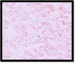
  • Post-mortem tissues were received from a one-day-old neonatal lamb that died despite antibiotic treatment overnight. The flock had been experiencing several cases of pneumonia-like symptoms in Swale lambs, from approximately eight hours to a few days of age. All had been bright but with increased respiratory rates initially. On histopathology, there was evidence of an interstitial pneumonia (Figure 1). Von Kossa stain highlighted some positive material in the alveoli which confirmed the presence of mineral, including but not limited to calcium containing compounds. An inhaled irritant was therefore a possible cause, including lime, but other causes were also possible, and it was advised that the findings were interpreted alongside clinical history/management.


Figure 1: Scattered intracellular and extracellular material in the lung of a lamb with interstitial pneumonia shows positive staining (black granules) with von Kossa special stain (mineral).

  • Lung samples were received from a four to six-week-old hill lamb, one of two to die suddenly in a flock vaccinated with multivalent clostridial-Pasteurella vaccine. Histopathology detected acute, fibrinous interstitial pneumonia with intralesional bacteria and intravascular fibrin thrombi consistent with a gram-negative bacterial septicaemia and terminal DIC, which would have accounted for the death of the Bibersteinia trehalosi was considered the most likely pathogen, but Mannheimia haemolytica, Pasteurella multocida and other gram-negative bacteria were also possible.
  • Infection with the lungworm species, Muellerius capillaris, was diagnosed on Baermann’s testing of a pooled faeces sample from four to six-year-old goats, one of which was coughing and had nasal discharge. Goats are known to be more susceptible to capillaris infection than sheep.
  • Infection with the lungworm species, Dictyocaulus filaria was diagnosed on Baermann’s testing of a pooled faeces sample from 12-month-old gimmer hogs in which there was coughing across the group of about 30 animals and increased lung sounds in the two animals examined. filaria is the most pathogenic lungworm species in sheep.

Gastrointestinal disease

  • Cryptosporidiosis was diagnosed in scouring lambs on a farm where one employee was reported to be unwell (though the clinical presentation and/or diagnosis for the person were not mentioned in the clinical history). Cryptosporidiosis is a known zoonotic disease and may have been implicated in the worker’s Infection typically occurs between 5 and 15 days of age and Cryptosporidium parvum, the protozoan parasite responsible, is transmitted by the faeco-oral route. The oocysts are resistant and widespread environmental contamination can occur from which lambs acquire infection.
  • Parasitic gastroenteritis was diagnosed on faecal egg counts in several flocks and goat herds, with presenting signs including ill thrift, scour and, in one lamb, rectal Raised pepsinogen levels consistent with parasitic abomasitis were also confirmed in some sheep.
  • Haemonchosis was confirmed in animals with high strongyle egg counts with a varying percentage of the eggs determined to be Haemonchus species using the Ovacyte method. In one case, two Suffolk ewes died suddenly over the weekend and, on PME, the viscera were pale and there was a large quantity of pleural and peritoneal 56% of a faecal strongyle egg count of 30,654 epg were determined to be Haemonchus species.
  • Among the cases of fasciolosis diagnosed this month was a sick six-year-old wether that was strongly positive on liver fluke coproantigen ELISA; interestingly liver enzymes in this sheep were not raised.
  • Among the several cases of Johne’s disease diagnosed this month on serology or faecal PCR was a two-year-old ram that had lost a lot of condition, had been scouring for a few months and had recently developed bottle jaw.

Skin and eye disease

  • Staphylococcus aureus was isolated from one of multiple abscesses found on a lamb. aureus may be associated with tick borne fever and subsequent tick pyaemia in lambs, so it was advised that the possibility of tick infestation/TBF could be considered.
  • Mycoplasma conjunctivae was detected by PCR on all three ocular swabs submitted from a flock in which many ewes were developing infectious keratoconjunctivitis.
  • Samples were received from the back of a hoof of a sheep (no history was provided). Histopathology detected two separate lesions in the samples. The first was consistent with a malignant neoplasm, most likely osteosarcoma. This is a rarity in large animal species and, when it does occur, it is usually found in the skull. Therefore, assuming it was osteosarcoma, the species and location were both very unusual. There was also evidence of parapox (orf) infection and secondary bacterial infection on the ulcerated surface. Local abrasion around the coronary band/lower limbs is likely to have predisposed to infection by the parapox virus leading to the localised lesions in this case.
  • Treponema medium and Treponema pedis were detected by PCR in swabs taken from an infected toe granuloma in a sheep. These treponemes are involved in the aetiology of CODD and toe granulomas are known to be a potential sequel of
  • Evidence of sheep scab exposure on serology was again found in flocks this month, signs including pruritus and wool slip.

Systemic and miscellaneous disease

  • urther cases of CLA were confirmed on culture and serology this
  • Exposure to Maedi visna virus was detected on serology in several
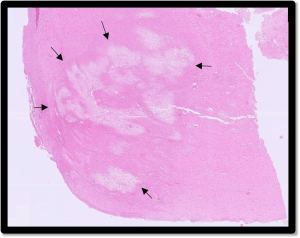
  • Pulpy kidney was suspected to be the cause of death in lambs from several flocks, with the detection of epsilon toxin on Clostridium perfringens toxin ELISA testing of ileal or caecal Brain histopathology is required to confirm the diagnosis, and this was achieved in two cases. In the first, a flock with a history of sudden death in two-week-old lambs, a lamb exhibiting neurological signs including paddling and tremors, was euthanased. Histopathology detected severe multifocal symmetrical widespread necrotising encephalopathy (symmetrical encephalomalacia) reflecting a vascular pathogenesis and typical of lesions seen secondary to epsilon toxin enterotoxaemia (Figure 2). Similar lesions were found in the brain of the second case, a three-week-old lamb in another flock, and this lamb also had multifocal acute renal tubular necrosis, also consistent with a diagnosis of pulpy kidney.

Figure 2: Areas of necrosis (arrows) in the brain of a lamb, suggestive of encephalomalacia as seen with Clostridium perfringens Type D epsilon toxin enterotoxaemia
  • Post-mortem tissues were received from a Mule ewe which had exhibited neurological The farmer had treated with penicillin/streptomycin, NSAIDs, vitamins B1 and B12, and calcium three weeks post lambing and Listeria was now suspected. This was confirmed on histopathology with the detection of severe, suppurative, granulomatous and plasmalymphocytic, necrotising, rhomboencephalitis and meningitis, consistent with listerial encephalitis due to Listeria monocytogenes.
  • Two four-year-old Cheviot ewes exhibiting neurological signs two to three weeks post lambing in a Devon flock were seropositive for Louping Ill virus on the HAI test, with evidence of IgM predominance supportive of recent infection.
  • Trueperella pyogenes and Helcococcus ovis were isolated from a joint swab from a Texel cross lamb in a flock with a high rate of with septic T. pyogenes is a known cause of pyogenic infections in livestock and H. ovis is an emerging pathogen in livestock that has been isolated from cases of septic arthritis in lambs and calves.
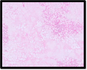
  • Postmortem tissues were received from a ewe that had been due to lamb in two Before death, it exhibited severe respiratory signs and pyrexia and, on PME, numerous hard caseous type lesions were seen throughout the abdomen. On histopathology there was extensive, marked, chronic, fat necrosis and saponification, suggesting a diagnosis of lipomatosis (Figure 3). This is not uncommon in cattle where it can occur in any part of the omental, mesenteric, and retroperitoneal fat. However, the condition is considered rare in sheep. The aetiology of lipomatosis is unclear; it can be an incidental finding or potentially cause problems such as obstructions of the gastrointestinal tract.

Figure 3: Fat necrosis and saponification in a sheep with hard lesions throughout the abdomen, suggestive of lipomatosis


Camelids

  • An adult alpaca with a history of inappetence and weight loss was found to have parasitic gastroenteritis (1000 epg strongyles) and fasciolosis (on fluke microscopy), with rumen fluke eggs also being found in the faeces, although the clinical significance of the last is unclear in South American A further case of parasitic gastroenteritis was diagnosed in a one-year-old alpaca with scour, condition loss and a strongyle egg count of 1600 epg.
  • Tissues were received from an ill thrifty seven-year-old alpaca with an infected mass behind the left mandible, which had only partially responded to antibiotics. On histopathology, two of the sections of the mass consisted of haired skin with non-specific peri-vascular dermatitis. In the third small section, there was a proliferation most suggestive of squamous cell carcinoma. This is a not uncommon neoplasm of camelids. In the skin, these tumours are usually locally invasive, but uncommonly metastatic until late in the course of the lesion. When spread occurs it is usually to the local lymph nodes in the first instance.
  • A lump on the lip of a female alpaca was found on histopathology to be a round cell tumour. Possible differentials included plasmacytoma, mast cell tumour, lymphoma, or a histiocytic neoplasm. It was not possible to state if the tumour was benign or malignant. It was suggested that immunohistochemistry could be used to obtain a more definitive diagnosis.
  • Post-mortem tissues were received from a 48-hour-old cria that was born five weeks prematurely. Histopathology detected severe, acute hepatic necrosis with intralesional gram-negative bacteria, suggesting terminal neonatal septicaemia. Given the presentation, colisepticaemia was considered the most likely, although other gram-negative bacteria (e.g. Salmonella spp, Pasteurella spp etc) were also possible.

Pigs

  • Tissues were received from an eight-year-old sow with a pedunculated mass protruding from the lips of the Histopathology was consistent with an inflamed endometrial polyp with cystic endometrial hyperplasia. A very small number of glandular structures showed some cellular atypia which could have been dysplastic change in the endometrial glands associated with adjacent inflammation. However, it did raise concern for malignant neoplasia (e.g. uterine adenocarcinoma).
  •  

Deficiencies and toxicities

  • Hypomagnesaemia was diagnosed on testing of two adult dairy cows which were showing signs of being ‘nervous’ when out grazing on lush pasture.
  • Five Aberdeen Angus cross suckler cows on one farm were in poor body They had been fed silage throughout the housing period with no added minerals, and no forage analysis had been carried out. No evidence of liver fluke infestation was found on faecal microscopy. Four of the five tested cows were hypocupraemic and all had low urea levels consistent with a low level of rumen degradable protein (RDP) in the diet.
  • Lead toxicity was diagnosed by testing of kidney tissue from a calf which was the fourth calf to die recently from the The animal had been found recumbent, seizuring and pyrexic and died a short time later. The kidney lead level was 20.98mg/kg FT. Lead levels of >2.0mg/kg FT in kidney are considered consistent with lead poisoning in cattle. Liver or kidney concentrations of >0.50 mg/kg FT indicate that undesirable lead residues are present that will exceed the permitted levels for offal as food. It was advised that the incident be reported as a potential food safety incident to APHA for the case to be managed to avoid contamination of the food chain
  • On one dairy farm cases of scour and pneumonia persisted in young calves despite evidence of excellent passive transfer and a clean, dry environment. Testing of calves at one month of age identified all five tested animals as having low Vitamin A levels and four of the five had evidence of Vitamin E deficiency.
  • Evidence of Zinc deficiency was identified on testing of a five-week-old Limousin bull calf which showed signs of sudden onset alopecia two weeks after The zinc level in serum was 9.5 µmol/l (RI=12.2-18.2umol/l). In another case of suspected Zinc deficiency, a four-year-old ewe with ill thrift and poor skin and fleece condition was found to have a zinc level of 3.8 µmol/l (RI= 7.7-16 µmol/l)
  •  

Follow us on Facebook

https://www.facebook.com/Axiomfarmvetlab/

This newsletter is sent by e-mail to each vet practice but if you would like a copy sent to your individual e-mail account please contact us at dsfarm@axiomvetlab.co.uk and we can add you to our circulation list.

Related Posts